

21-07-2013 14:49
Marja PennanenHello, these pinkish ones are up to 0,5 mm wide.Th
23-07-2013 00:00
 Malcolm Greaves
This group of small Scutellinia (maxium size 2.5mm


21-07-2013 14:57
Marja PennanenThese are about 0,2 mm wide.The spores are about 1
23-07-2013 07:25
 Zuzana Sochorová (Egertová)
Good morning,could anyone send me this article?Svr
20-07-2013 19:25
 Joop van der Lee
Found on horse dung.I have looked into the differe
Here's already my next problem:
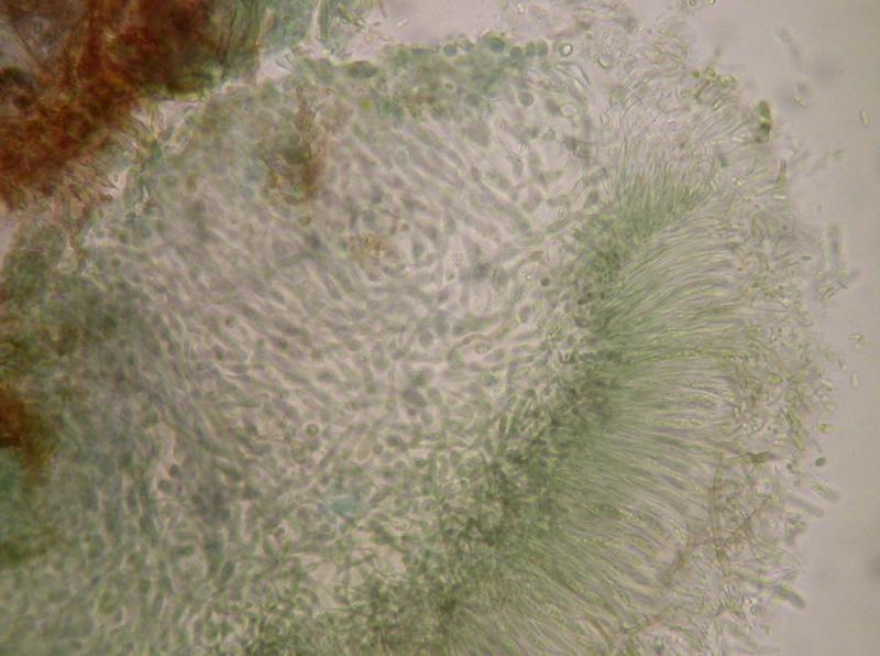
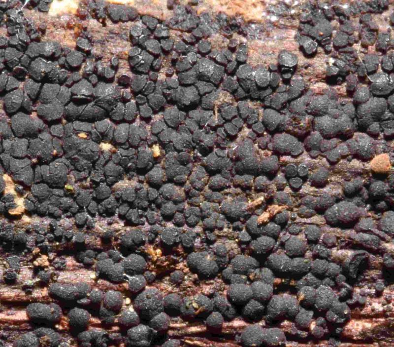
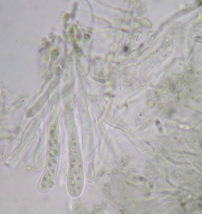
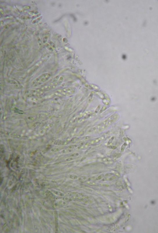
Substrate: (Yet) Unidentified deciduous wood
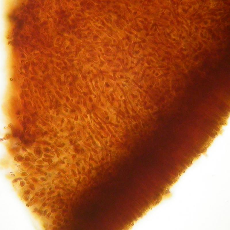
Macro: Apothecia, up to 0.5 mm diam. (as single apothecia), exceeding 1 mm in confluent parts. Densely aggregated and often more or less coalescing to confluent parts. Black, with a distinct margin when young, then flat without margin and later convex. Exuding as a red-brown colour in KOH.
Micro: The whole disc is incrusted with a brown substance and structures are difficult to see when prepared in water. This Substance dissolves in KOH as a reddish exsudate and then the structures are hyaline to distinctly green. Asci 35-40 x 5 µm, IKI-, Melzer-, Spores hyaline, 4.5-5.5 x 1.2-1.6 µm, with two oildrops, aseptate. Paraphyses about 1 µm diameter. Excipulum not clearly differentiated from medulla, the medulla consisting of gelatinous tissue with loosely intertwined, irregularly expanded hyphae (Photos).
Thank you for any help
Stefan

Ist was Durelloides.
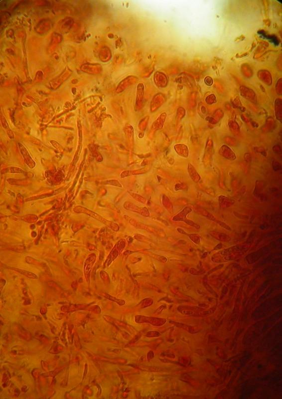
Hier noch zwei Fotos von Sporen/Asci
LG
Stefan
danke, das hilft! Vergleiche mal meine Zeichnungen im Verzeichnis Phaeangella = Durella redbrown, und da "bigutt spores narrow ionom". Da sind zwei Funde, einer aus USA, der andere aus Luxemburg. Die wuchsen aber an ansitzenden Ästen.
Hast du keine Skala in deinen Fotos? Dann könnte ich etwas nachmessen.
Das Auflösen und Austreten des rotbraunen Pigments ist typisch für diese Art (ionomidotisch). Irgendwie erinnert er auch an Ionomitodis fulvotingens, aber nur mikroskopisch inklusive Excipulum.
Grüße
Zotto
Habe hier mal noch einen 10 mü Massstab reingeflickt. Was besseres habe ich im Moment nicht. Hilft wohl nicht viel. Kann aber gerne nochmals gewisse Sachen nachmessen...
LG
Stefan
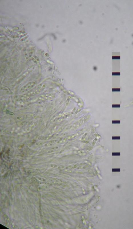
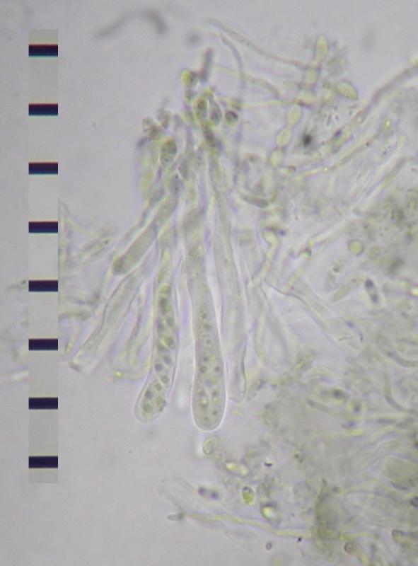
Asci tot 45 x 4.5-5.5 µm,
Sporen etwa 4,5-5,5 x 1,8-2 µm