05-05-2026 22:40
Gernot FriebesHi,I believe this is a Plagiostoma growing on a Sa
04-05-2026 18:13
 Stephen Martin Mifsud
ID request for what seems to be a true aquatic fun
04-05-2026 16:39
Stephen Martin Mifsud
ID request: This specimen was collected in Malta o
28-07-2011 18:31
 Alex Akulov
Dear FriendsToday I made the pdf file of Velenovsk
28-04-2026 20:07
 Lothar Krieglsteiner
... on twig in the air at standing Ceratonia siliq
04-05-2026 09:50
 Castillo Joseba
Me mandan el material seco de Galicia,(España) re
02-05-2026 12:42
Alain BRISSARDBonjour à tousJeuidi 30 avril dernier on m'a remi
02-05-2026 13:06
Pauline. PennaBonjour Please can someone help me with this id
01-05-2026 22:45
 Thierry Blondelle
Bonjour à tous, Une récolte sur bouse séchée d
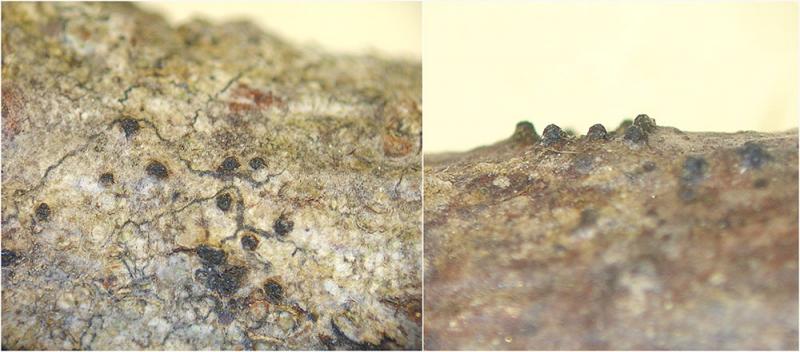
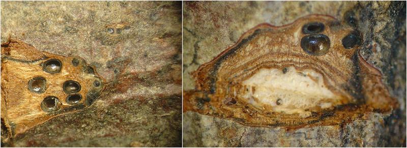
I have Collected a Diatrypaceae on hardwood.
Here are some features:
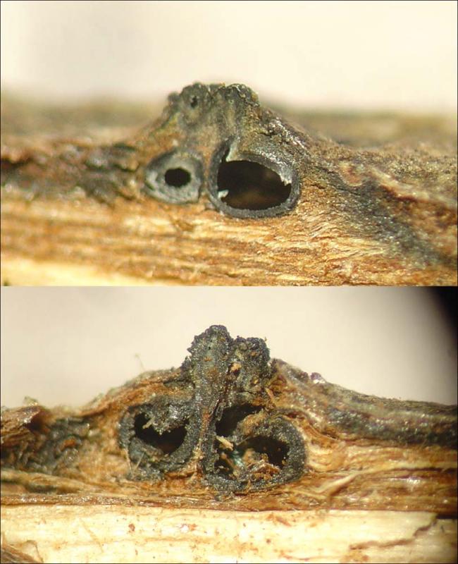
Perithecia immersed, usually separated, spherical / ovoid with a diameter of 600-900um.
Ostioles emerging separately. With a round shape the diameter is about 350um.
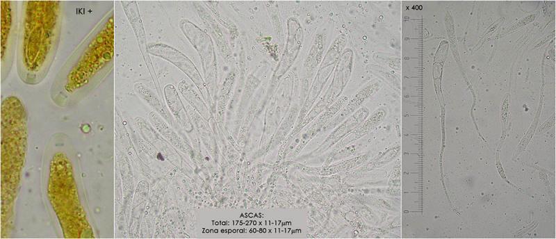
Asci octosporadas on a long pedicel clearly IKI +
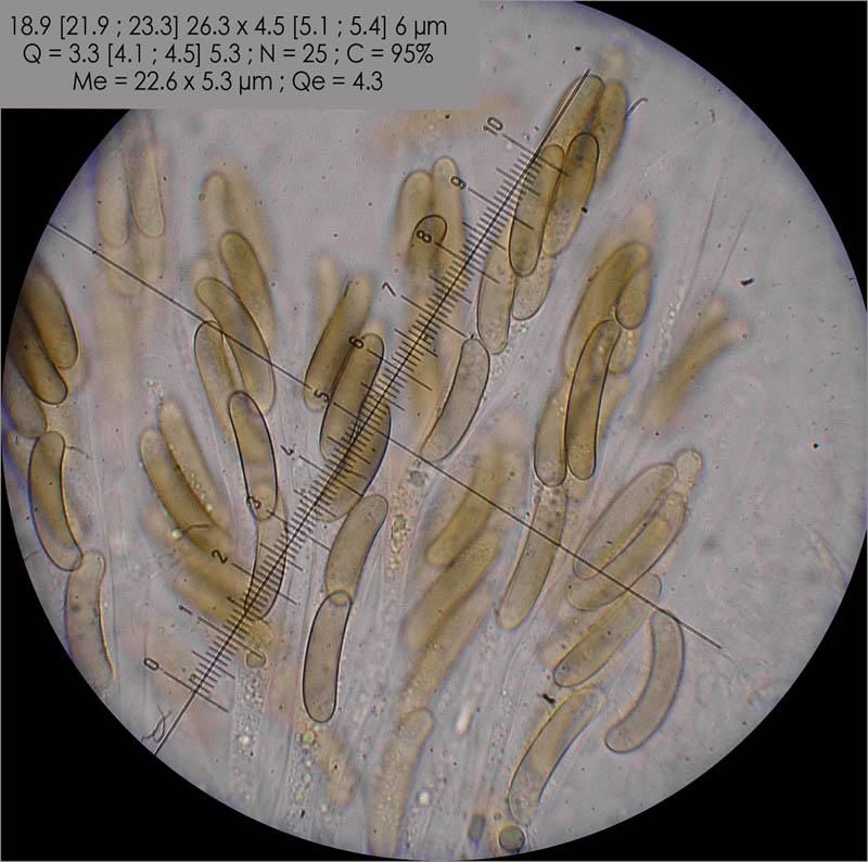
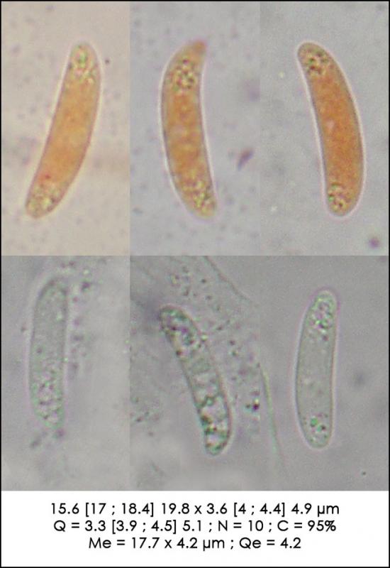
Are not mature perithecia and spores are difficult to obtain, however, the size of which are measured are on average 17.7x4.2um, and are slightly allantois.
With these data, I think it might be Cryptosphaeria and within this genre fits better with C.subcutanea.
You can give me your opinion?
Thank you very much, greetings
Susana

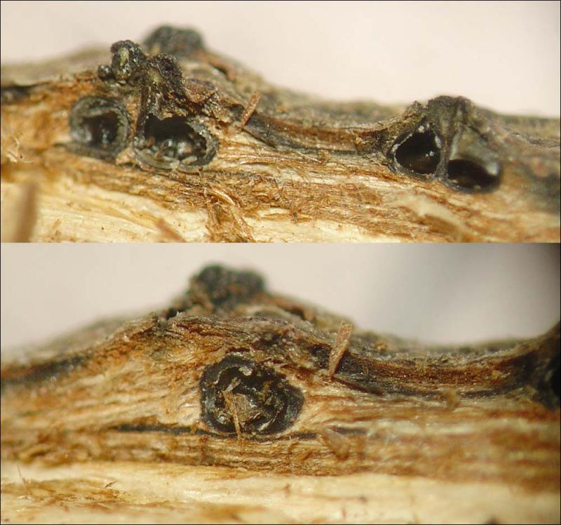
again an interesting find. I agree it resembles Cryptosphaeria by many traits but in this genus the wood surface beneath the stromata is typically strongly blackened unlike what shows your photo 2.
I suggest you observe a stroma in vertical section and compare it to Rappaz' description.
Moreover, C. subcutanea which might fit as to ascospore dimensions differs in having inamyloid asci and a fairly boreal distribution on Salix.
Try to find mature ascospores to check whether they become pigmented or stay hyaline.
Good luck!
Jacques
I found mature perithecia, and spores are pigmented, they are brown.
I made cross sections, and I noticed that the perithecia are grouped two to three and their necks are emerging together.
There is a black dorsal line (Fig.2) and in some cases a ventral black line also appears (Figure 3).
Now I think it may be Eutypella dissepta. It can be?
Saludos
Susana