
27-04-2026 20:52
 Lothar Krieglsteiner
Found on hanging tiwg of Olea europaea in dried-ou
27-04-2026 18:48
Tony MoverleyCollected 23rd April 2026, Norfolk, EnglandSwarms
27-04-2026 17:41
Lothar Krieglsteiner
.. Algarve, same leaf than the last post. The con
27-04-2026 18:05
Lothar Krieglsteiner
... still attached at standing tree. The green con
27-04-2026 17:16
Lothar Krieglsteiner
.. Algarve, moist lying.The conidiomata look like
27-04-2026 12:54
Steve ClementsBonjour. Ce petit champignon blanc résupiné et
27-04-2026 09:59
Pauline. PennaBonjour Can anyone advise me on these pycnidia fo

22-04-2026 20:54
 Enrique Rubio
Hi to everybody.This Pyrenopeziza grew in moist le
Microscopic ascomycete.
Josep Torres,
24-09-2025 09:22
 Hello.
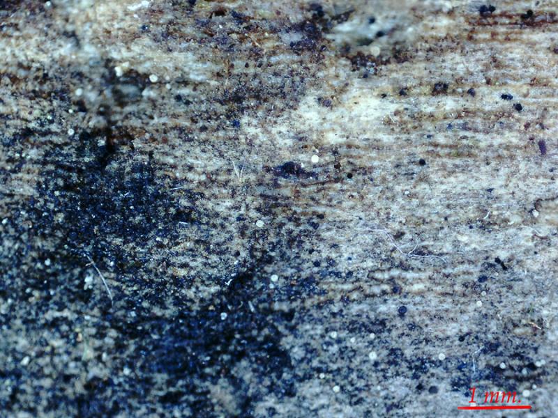
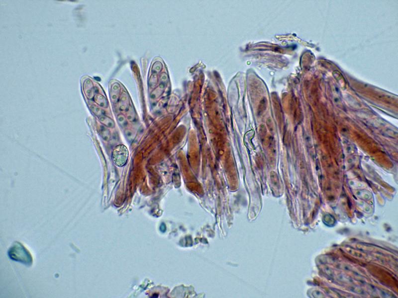
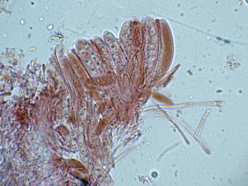
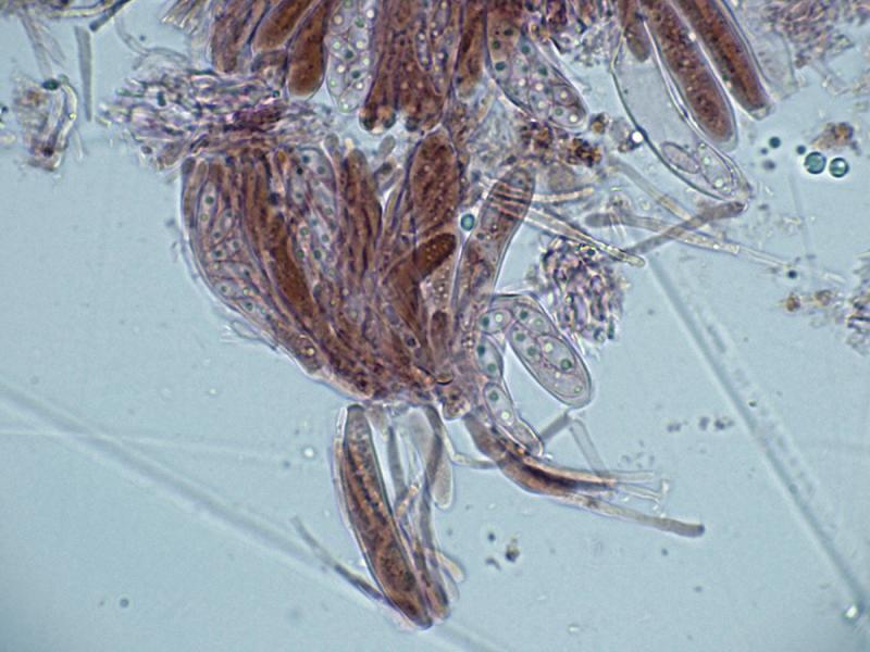
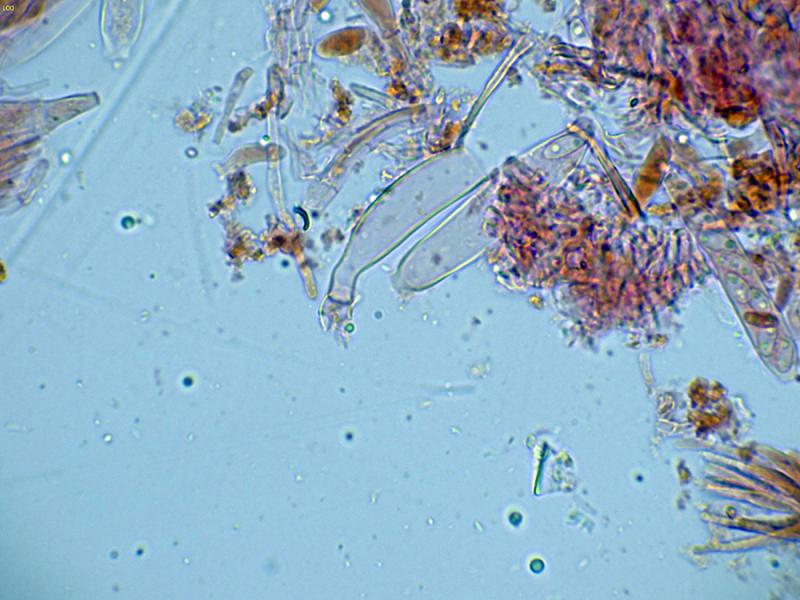
Very tiny apothecia, sprouting in a scattered manner, located on the surface of a semi-submerged trunk of Fraxinus sp.
Measuring only 0.07 to 0.11 mm in diameter, they are whitish in color and have a slightly hairy outer edge.
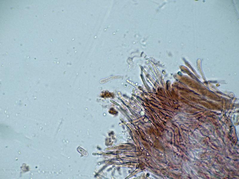
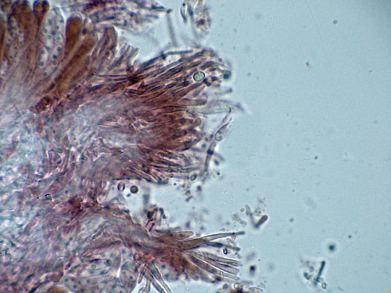
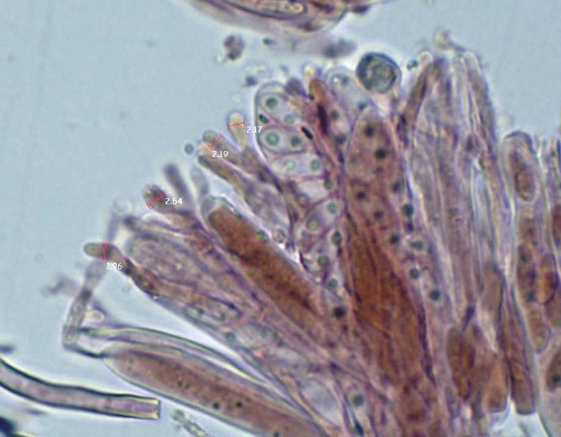
Marginal hairs with moderately pointed ends and inlaid surfaces.
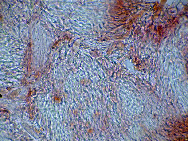
The medullary exciple hyphae are intricate, and below these, hyphae are already tending toward globose angularis.
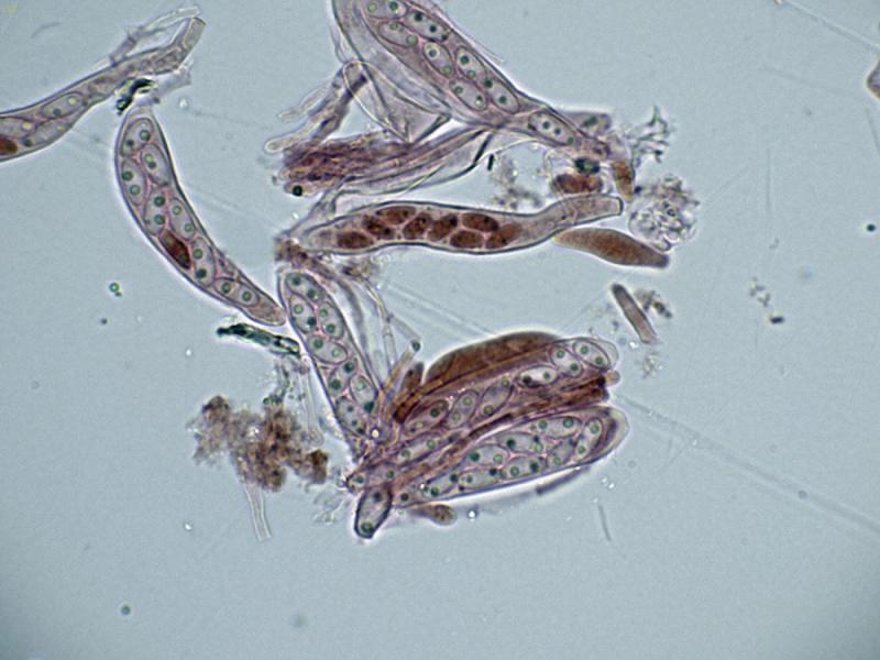
Octosporic asci, apparently without uncinules at their bases, mostly biseriate, and measuring (35.7) 40.9 - 52 (52.4) × (7) 7.7 - 9.1 (9.6) µm.
I can't provide data on the behavior of the asci to the Melzer test because I only managed to prepare a single sample with a single apothecium. The next day, the remaining apothecia had literally disappeared after dehydration.
Ellipsoidal ascospores with two large lipid droplets at the poles, with measurements of mature ascospores within the ascus of:
(5.9) 6.6 - 8.9 (9.3) × (2.9) 3.2 - 4.3 (4.7) µm
Q = (1.5) 1.8 - 2.4 (2.8) ; N = 30
Me = 7.6 × 3.6 µm; Qe = 2.1
The paraphyses are filiform, slightly or not at all widened at the apex, not protruding above the level of the asci, with one or two septa and a width of 2 to 2.55 microns.
Considering their characteristics, I think they are likely a Hyaloscypha, and among them is a Hyaloscypha microscopica, which, although the name would be appropriate, I have been unable to find any information about the species.
Any feedback from you would be most welcome.
Thank you in advance.
Best regards.
Hans-Otto Baral,
24-09-2025 10:17

Re : Microscopic ascomycete.
Will be difficult without the iodine reaction. Perhaps Hyaloscypha intacta which has inamyloid asci (in Lugol).
H. microscopica was not ever redescribed. SVrcek 1985 only combined it in Discocistella and Huhtinen 1990 did not study the sparse material.
I did not look up Velenovsky but assume that the hairs are not tapering.
Josep Torres,
24-09-2025 14:20
Re : Microscopic ascomycete.
Thanks, Zotto.
Considering its tiny size, Hyaloscypha intacta seems like a good option. This has been mentioned here before. Here's a link to a study very similar to my proposal, which you yourself considered Hyaloscypha intacta.
http://ascofrance.com/forum/16979/hyaloscypha-hyalina#
Best regards.
Considering its tiny size, Hyaloscypha intacta seems like a good option. This has been mentioned here before. Here's a link to a study very similar to my proposal, which you yourself considered Hyaloscypha intacta.
http://ascofrance.com/forum/16979/hyaloscypha-hyalina#
Best regards.