
12-03-2015 19:10
Steve ClementsHi,I tried to key out this minute asco using Ellis
13-03-2015 08:48
Katarina PastircakovaHello,if anyone has access to Butin, H. European
11-03-2015 19:06
 Jenny Seawright
Hello all, I was wondering if anyone might have a

10-03-2015 14:50
Jenny Seawright
Hello all, I noticed a gelatinous substance on con
04-03-2015 09:45
 Miguel Ángel Ribes
Hello friendsThis small, < 1 cm Peziza, was gro

20-02-2015 00:25
Miguel Ángel Ribes
Good night againThis dark-violet, 4-5 mm Marcellei
11-03-2015 13:20
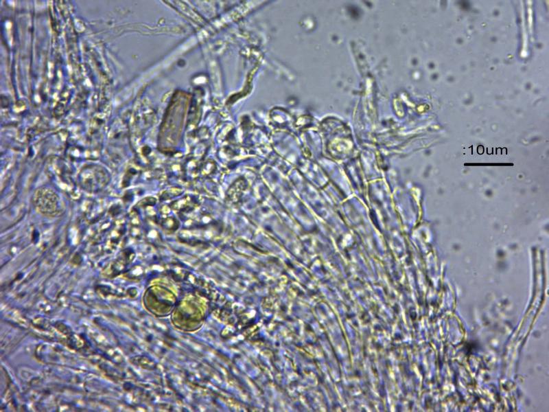
Chris JohnsonGreeting allFound on Bos taurus. Apothecia no more
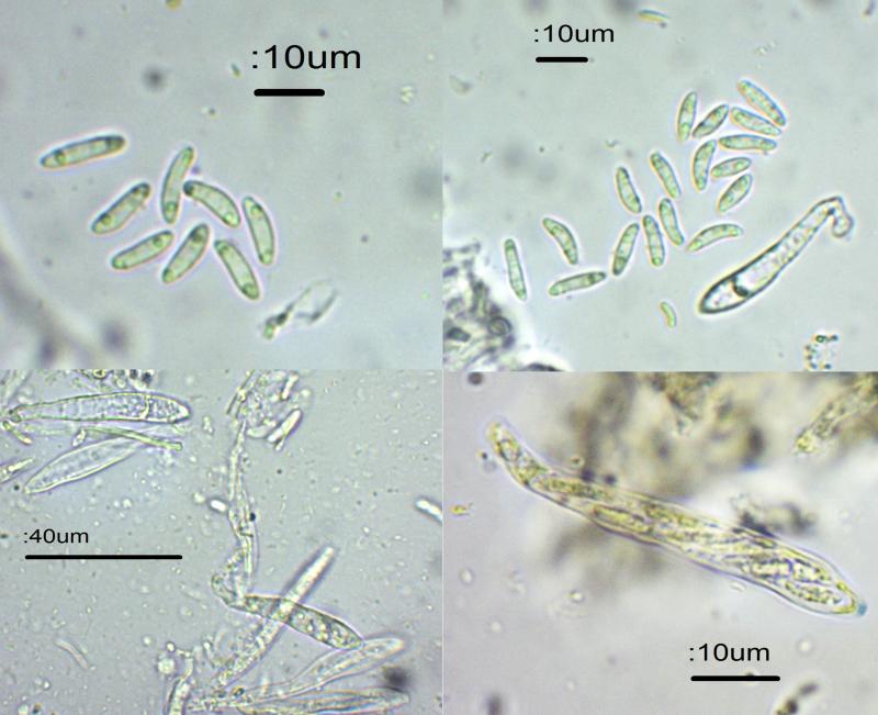
I tried to key out this minute asco using Ellis and Ellis and Fungi of Swizerland and got nowhere. So I resorted to scanning all of Peter Thompson's pictures in Ascomycetes in Colour with spore diagrams which looked right. The only one I was able to match up was Paraorbiliopsis minuta. Because Peter's descriptions are brief, I wasn't over-confident with the ID. The specimen was found on a stick under larch in a mixed wood, so may not be larch – it's an increasingly disturbed woodland right on the edge of Sheffield. The semi-transparent cushion-saped sessile fruit bodies were up to 350 mu (0.35 mm) wide so very small indeed. Colour pale yellow. I wasn't convinced that they were on a subiculum. On microscopy the asci discharged spores – an amazing sight. Hyaline spores with 2 guttules, 9-13.5 x 3-3.5, slightly curved. Asci about 50 x 6-7, tips bluing in Melzer's, inoperculate I think. Paraphyses hard to see but look to be thread-like.
Any comments would be very welcome,
Cheers,
Steve


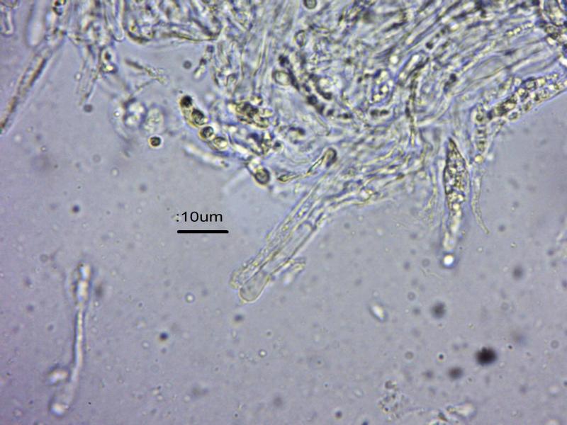
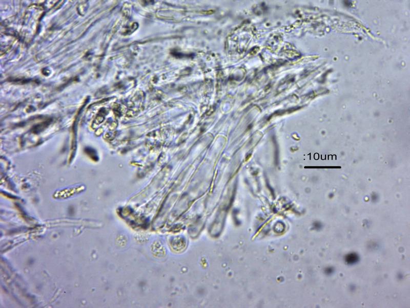
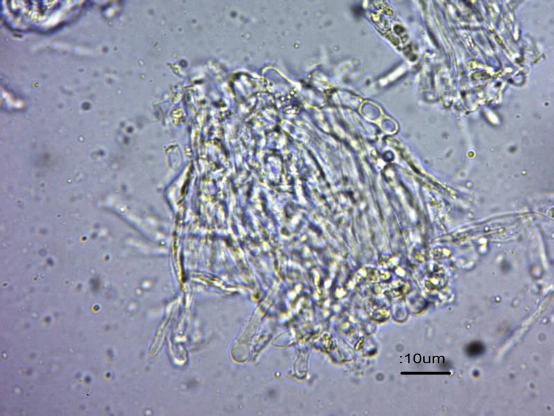
P. minuta is excluded because it is inamyloid. What you can do is looking in a squash mount on the margin from exterior (!), so put an apo upside down in the water drop and place the slide over it. Then you can try to look if there are any short hairs at the margin or the mid flanks of the excipulum. I suspect a Hyaloscypha. The genus Parorbiliopsis is undoubtedly nothing but a Hyaloscypha without hairs. Species with amyloid asci generally have tapered hairs, partly rather short and easily overlooked.
Zotto
(Peter Thompson has Paraorbiliopsis as "blue reaction with iodine" - is this an error?
Steve

at this level I really wouldn't be looking at Peter Thompson's book for further information; the drawings there are, unfortunately, a bit basic; I'll email you some useful stuff re Hyaloscyphaceae - and do look at Zotto's files at https://www.cubbyusercontent.com/pl/CC%2bAscomycota/_1354d48ffaad4b59bd3ffdbb35915d1f#CC%20Ascomycota/7a%20Helotiales > Helotiales > Hyaloscyphaceae without VBs
regards
Chris
No idea.....

Hi Steve, and everyone,
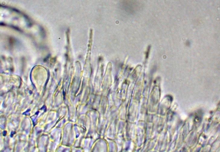
We lately had the very same question on our local SMMA forum a few days ago. A Parorbiliopsis collection was suspected for a tiny disco apparently whithout hairs and negative IKI reaction.
As I had the opportunity to receive and study it living, I discovered unconspicuous hairs not longer than 20 µm long as seen on the attached picture.
I concluded (with the other features, including positive IKI reaction.. in H. albohyalina var. spiralis)
Amitiés
Michel
do you mean your fungus had amyloid asci or not?
If not then such short hairs would point to H. intacta.
Zotto
That's very kind of you,
Steve
many thanks everyone for the assistance.
I shall record this as Hyaloscyphus sp., which is a new record for the Sheffield wood I am currently surveying,
Steve
Yes Zotto, the first try had showed unamyloid asci , probably due to too old Lugol.
(It was the opportunity to see that n your files the difference is light between H. intacta and the paorbiliopsis sp.)
Michel