24-01-2011 21:26
 Rodríguez Borja
Hi! Here´s a Acomycete from Madrid(Spain) b

23-01-2011 21:42
Gernot FriebesHi, I think this collection is close to Lasiosp

19-01-2011 19:52
 Björn Wergen
Hello again, I have recently found another inte

19-01-2011 17:36
René DougoudChers Tous, Je recherche la publication de Cibo
Red Lamprospora from Spain
Rodríguez Borja,
24-01-2011 21:26
 Hi!
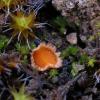
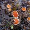
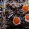
Hi!Here´s a Acomycete from Madrid(Spain) between moss. 23-I-2011
I´ve thought about a species of Lamprospora but I cannot find something like..
They are red disc around 0.2 cm. at the centre red and in the margin pale orange.
The microscopic caracteristics:
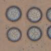
Spores globose uniguttulate, with reticulum formed by ridges wich are >1µm high and 0.8 wide.
15.2 [16.6 ; 17.6] 19.1 x 13.3 [14.2 ; 14.8] 15.6 µm
Q = 1.1 [1.2] 1.3 ; N = 15 ; C = 95%
Me = 17.1 x 14.5 µm ; Qe = 1.2
Ascus around 180µm, paraphyses as the ascus and subcapitate 4-5 µm.
Also the are few hairs of 150µm.
Thank you very much!
Rodríguez Borja,
24-01-2011 21:29

Re:Red Lamprospora from Spain
Photo sorry
Rodríguez Borja,
24-01-2011 21:32

Re:Red Lamprospora from Spain
Here is the photo I cannjot up as a normal text. Its a document
Raúl Tena Lahoz,
24-01-2011 21:55

Re:Red Lamprospora from Spain
Hola Borja
Podría tratarse de Lamprospora tortulae-ruralis Benkert. Adjunto mis macro y esporas al micro de esta especie. Aunque la ornamentación es muy discreta en esta especie. Sale sobre Tortula ruralis. La imagen macro de abajo a la izquierda parece de Tortula ruralis. La macro cuadra bien con el amplio margen. Observo alguna espora subglobosa en la micro, ¿son habituales?
Espera a alguna otra opinión. A lo mejor Gibert Moyne o Enrique Rubio o ...
Un abrazo,
Raúl
Maybe it can be Lamprospora tortulae-ruralis Benkert. I attach my macrophotos and the spores seen under micro. The ornamentation is rather low in this species. It grows on Tortula ruralis. In the lower left macro image it seems to be Tortula ruralis. The macro pics recall this species because of the broad margin. I see some subglobose spores in the micro, are they usual?
Wait for more opinions. Maybe Gilbert Moyne or Enrique Rubio or ...
Raúl
Rodríguez Borja,
25-01-2011 22:17

Re:Red Lamprospora from Spain
Hola Raúl como bien dices estaba sobre Tortula sp. Es un género que es la primera vez que tengo entre las manos y la verdad no tengo mucha idea.
Por la microscopia que veo con la tuya coincide bien pero tengo una pregunta, el reticulo de la mía era muy regular y en tus fotos lo veo bastante irregular, eso tiene algún caracter determinativo?
Tu determinacion va muy bien, si nadie aporta nada nuevo lo dejo como Lamprospora tortulae-ruralis.
Te agradecería si me pudieras pasar algo de informacion de tu especie o del género en general.
Borja Rodríguez
Hi Raul as you say was on Tortula sp. It is a genre that is the first time I have in my hands and I have not really great idea.
By microscopy to see, yours fits well but I have a question, the reticulum of mine was very regular in your photos I see quite irregular, it has a certain character?
Your determination is fine, if no one brings anything new I leave it like Lamprospora tortulae-ruralis.
I'd appreciate if I could pass some information of your species or genus in general.
Borja Rodríguez
Gilbert MOYNE,
26-01-2011 15:00
Re:Red Lamprospora from Spain
Bonjour,
Je pense qu'il s'agit bien de Lamprospora tortulae-ruralis Benkert bien caractérisée par son habitat sur Tortula ruralis, sa marge claire et abondante et son ornementation sporale faite d'un réseau aux mailles très irrégulières (des grandes et des petites).
Amitiés
Gilbert
Je pense qu'il s'agit bien de Lamprospora tortulae-ruralis Benkert bien caractérisée par son habitat sur Tortula ruralis, sa marge claire et abondante et son ornementation sporale faite d'un réseau aux mailles très irrégulières (des grandes et des petites).
Amitiés
Gilbert
Rodríguez Borja,
26-01-2011 22:55

Re:Red Lamprospora from Spain
Thank you very much Gilbert and Raúl again. So, I file as Lamprospora tortulae-ruralis Benkert
greetings
borja Rodríguez
greetings
borja Rodríguez

 14048.jpg


