23-01-2017 18:02
 Enrique Rubio
Hi to allI would like to know your opinion on thes
21-11-2017 11:59
Peter ThompsonHello Everyone,I have found a hyphomycete which gr
04-11-2017 21:56
Thorben HülsewigHi there,today i found this anamorph fungus that g
18-11-2017 07:44
 Andrés Valverde Valera
Bonjour, Je ne sais pas si peut être cette espèc
18-11-2017 21:19
 Edmond POINTE
Bonjour,Je suuis perplexe quand a mettre un nom su
18-11-2017 14:26
 Bernard Declercq
Dear forum members,Who can supply me a pdf of foll
Peziza
hannie wijers,
21-11-2017 17:38
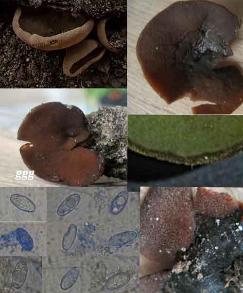
It's a while ago since I was her for the last tiime. But today I would be pleased with some help from you. I'm struggling already a fewe days with this peziza.
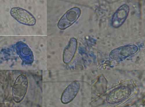
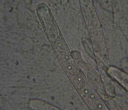
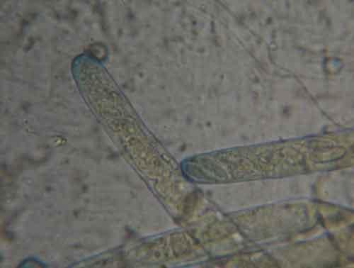
A friend brought along a peziza. The frb grow underneath or on very decaying wood that lay on the earth. We don't know what kind of wood.. It concerns mixed forest The small ones frb are about 1,5 cm till about 4 cm large. The outside of the asco is dark brown and a bit flaky, the inside is olive green and on the outside edge there seems to be a brown edge. Asci 300 x 15 ?m, spores 15-19 x 8-9 ?m. Mature spores are warty and on some tracks seems to be a network. .
Asci J+. Is this possible Peziza badia?
I hope one of you can help me with these information
Nicolas VAN VOOREN,
22-11-2017 19:17

Re : Peziza
Is it possible to see the microscopic pictures in a larger size?
hannie wijers,
22-11-2017 20:13
Re : Peziza
Hello Nicolas,
here larger drom spores and asci. Is this better toe see. Thank you
groetenHannie
here larger drom spores and asci. Is this better toe see. Thank you
groetenHannie