21-07-2025 19:22
Ethan CrensonHello all, Here is an Orbilia found by a friend

14-07-2025 11:20
 Michel Hairaud
Bonjour, Voici une espèce de (?) Hyaloscyphace

18-07-2025 23:03
Hello.Fruitings between 51 and 130 microns in tota

16-07-2025 17:34
 Bernard Declercq
Hello,I have trouble distinguishing above mention

16-01-2023 21:31
 Riet van Oosten
Hello, Nearby the find of Calycina claroflava on

14-07-2025 17:55
Yanick BOULANGERBonjourAutre dossier laissé en suspendJe viens de

14-07-2025 11:17
Yanick BOULANGERBonjourJ'ai un dossier Jackrogersella qui est rest

14-07-2025 15:52
Gernot FriebesHi,I wanted to share this collection on Rubus idae
Orbilia nemaspora ...or aurantiorubra?
Ethan Crenson,
21-07-2025 19:22
Hello all,
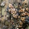
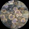
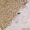
Here is an Orbilia found by a friend in a wooded NYC park on a fallen, well-rotted hardwood branch. The apothecia are a fairly dark orange color with a slightly crenulate margin, however, in micro there are no glassy processes at the margin, only globose cells.
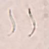
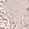
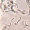
Spores are narrow, fusiform, curved in two directions (helicoid...) with the spore body at the acute end, the tail-end more rounded. Spores are 8.1-9.1 x 0.6-0.9µm measured "straight" rather than following the curved length of the spore.
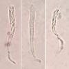
Asci are truncate when viewed from a particular direction. Spores woven together inside, usually the four top spores with the SBs oriented upward in the asci. Measurements, 36-37 x 3.5µm (though at this point I have not measured more than 2 asci).
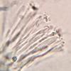
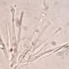
Paraphyses sometimes narrow, but also with somewhat swollen apices, around 2µm wide. The paraphyses held together with a substance that looks somewhat granular (? ... my photographs are not great on this detail).
My theory, currently, is Orbilia nemaspora, although I am also considering O. aurantiorubra. Any input would be appreciated.
Thanks in advance!
Ethan
Hans-Otto Baral,
21-07-2025 20:24

Re : Orbilia nemaspora ...or aurantiorubra?
The margin is not hit by our section, so how can you be sure? But I remember nemaspora without glassy processes. The spores are surely curved in only one direction, otherwise they would not fit in the bundle. On photos it is qiote impossible to decide the direction.
O. aurantiorubra is completely excluded, its spores being by far less curved. Yes, O. nemaspora is a good hit.
Ethan Crenson,
21-07-2025 20:44
Re : Orbilia nemaspora ...or aurantiorubra?
Hi Zotto! Thank you for your kind reply.
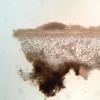
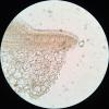
No, the photos in section excluded the margin (the section broke). The 5th photo shows the marginal cells from above in a squash. Does that not contain the information needed?
In the spore photo, the spore in the center has a widening, almost a bulb at the bottom end. I interpreted that as the end of the spore curving toward the lens, creating the illusion of that end getting larger.
I have uploaded a couple of new photos that may show the dimensionality of the spores a bit better.
I also have a short video https://youtu.be/CJ_rw62i8-k
Hopefully that will suffice. Apologies that my documentation is poor today.
E
Hans-Otto Baral,
21-07-2025 21:05

Re : Orbilia nemaspora ...or aurantiorubra?
I thought that was also a section. The video is very nice but only you know if you moved your objective up- or downwards. When knowíng this one can say if it is a righthand thread (which I assume).
I see the basal end poiinted, not bulbous.
Ethan Crenson,
21-07-2025 21:10
Re : Orbilia nemaspora ...or aurantiorubra?
But still O. nemaspora in your opinion?
Hans-Otto Baral,
21-07-2025 21:27

Re : Orbilia nemaspora ...or aurantiorubra?
Yes.

