05-06-2026 11:02
Thomas Læssøehttps://svampe.databasen.org/observations/10596691
07-06-2026 15:10
 William Slosse
Hello everyone,On 05-06-26, I found following asco
07-06-2026 12:43
Steve ClementsBojour. This was a strange find on a stick on my
07-06-2026 12:09
 François Freléchoux
Bonjour, Voici une brève description de ce qui m
12-07-2015 00:05
 Nedim Jukic
This one from the same locality as the previous on
06-06-2026 17:44
Steve ClementsBonjour, This disco was on planed wood 3 x 1.5 cm
14-08-2016 23:15
 Alex Akulov
Dear friendsCan you help me to find the descriptio

04-06-2026 11:36
Gernot FriebesHi,found on Vaccinium myrtillus.Asci: IKI –, 8-s
05-06-2026 12:10
François Freléchoux
Capitotricha sp. sur Lonicea caerulea Caractères
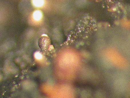
It occurs on exudate of wounds of Prunus pensylvanica in PEI and NB Canada.
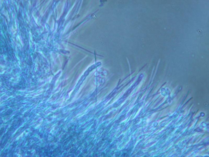
The ascomata are about 200-400 um tall and 150-200 um wide, and urceolate in shape. The walls are light brown and become melanized as it matures. Internally, the walls consist of linear, periclinally arranged hyphae. The asci are narrowly cylindric (c. 35 x 4 um), and lack an evident apical apparatus. They break down at maturity, producing a dry mass of spores that collects around the ostiole, and within the cavity in the upper part of the ascoma. Externally, the spore mass appears white to very pale yellow. The ascospores are uniseriately arranged, 8 per ascus, colourless, unornamented, and c. 3-4 x 2-3 um. They are ellipsoidal, but slightly compressed on the long axis.