30-05-2026 21:12
Philippe PELLICIERSur branche de mélèze (Larix) près de la neige,
31-05-2026 10:35
Hulda Caroline HolteHello,I collected this species growing on a rather
25-05-2026 16:35
 Bernard CLESSE
Bonjour à toutes et tous,J'ai trouvé récemment,
29-05-2026 15:35
daniel FERREBonjour à tous,Je voudrais votre aide pour cette
28-05-2026 16:15
James MitchellHello,Does anyone have the original publication of
28-05-2026 11:06
Thomas Læssøehttps://svampe.databasen.org/observations/10596750
23-05-2026 11:44
 Charles Grapinet
Hello, I am having trouble identifying this copro
25-05-2026 16:44
François BartholomeeusenHi forum members,During an excursion organised by
26-05-2026 21:25
Dirk GerstnerHello everyone, I'm completely stumped by this li
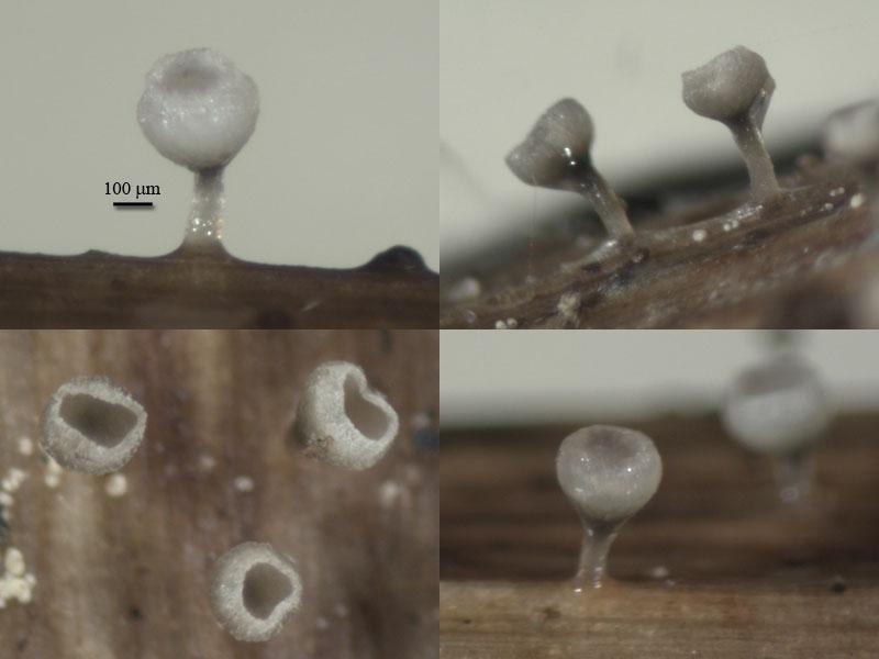
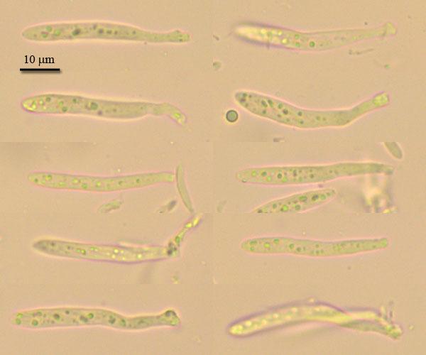
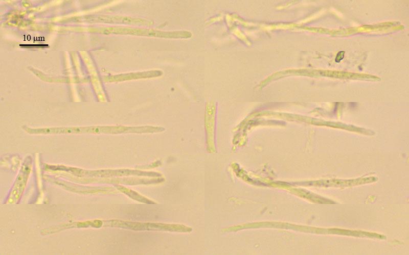
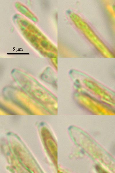
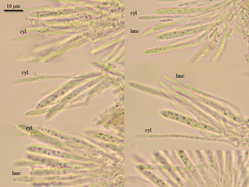
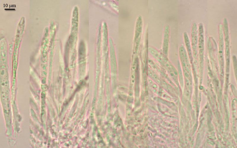
Apothecia goblet-shaped, receptacle deep-cupulate, to 0,5 mm in diam, stipe thin (100 mk), the same high as cup, all frb up to 1 mm high; stipe brownish, translucent, receptacle brownish at base, lighter to white at margin (when dry edge powdery from incrustation), edge rised under hymenium surface forming narrow collar.
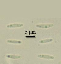
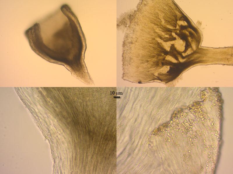
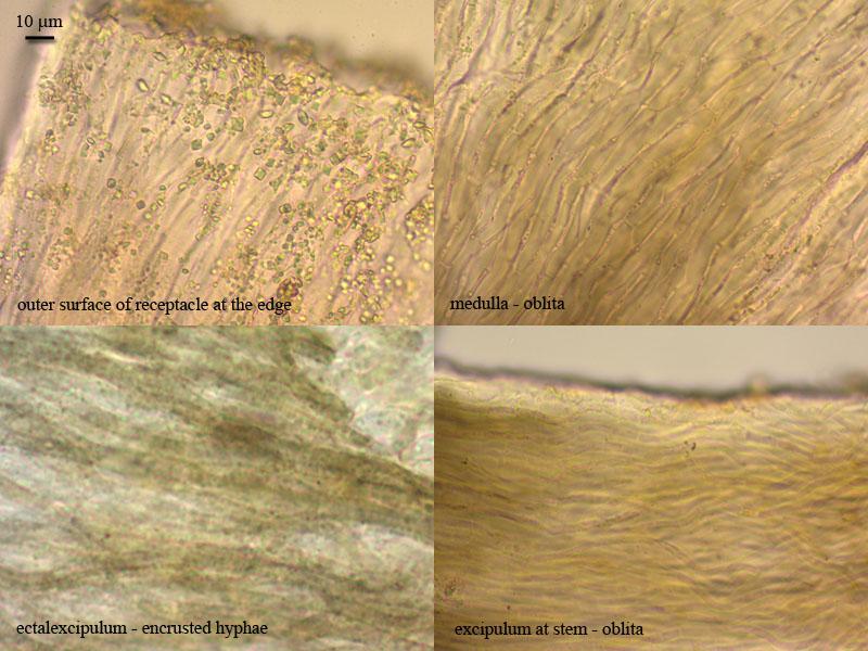
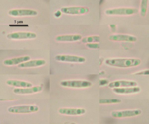
Excipulum from textura oblita, but outer layer of receptacle formed by porrecta, hyphae with rough walls (brown); margin from textura oblita, with abundant crystals; asci clavate, with crozier, with small euamyloid pore, 33,5-43 x 4,2-5,2; paraphyses lanceolate (not clear difference in two types), septate at base, slightly exceeding the asci, up to 3 mk broad in largest part; spores narrow-ellipsoid, with small guttules, 8 (7-9,4) x 1,7 (1,5-2,2) (N=18).
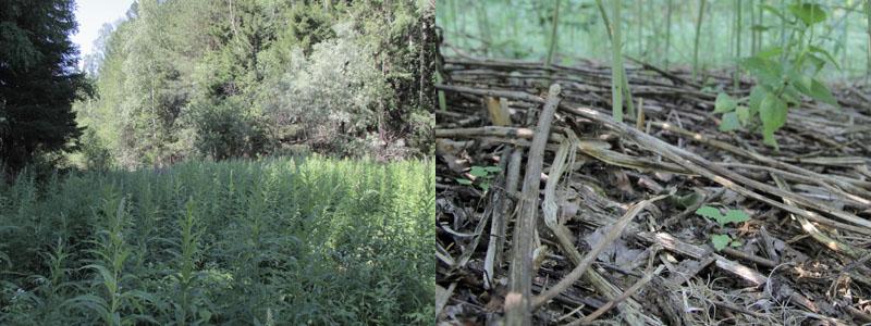
On dead stems of Glyceria triflora at forest edge, N61,090492 E69,480253, 26.06.2012.

You do not have any micropics in vital state? Here I suspect multiguttulate paraphyses as typical of Cyathicula.
Useful should also bi a photo of the apical rings in IKI. If you compare their shape with those of Crocicreas gramineum, we could perhaps see a distinct difference.
You say paraphyses lanceolate, but I see also cylindrical ones.
Zotto
i will send you vital photo,
there are pictures of ring, it it differrent.
right, i was confused with paraphyses, they were badly seen in previous specimen. Since all hymenial parts smaller, differences not so clear. But now i checked again and think there are also two types, lanceolate and narrow (these rarely seen).
i am not sure about VBs since lack of experience seing them in vital, but what would you say?
On your spore photo I think that two spores are alive (lower left, central right). You say KOH, is this true for all spore photos?
I will compare with Cyathicula starbaeckii.