10-06-2026 12:54
Steve ClementsBonjour encore, Pouvez-vous m'aider, s'il vous pl
09-06-2026 18:32
Camille MertensSur morceau de roseau immergé 0,5 - 0,7 mm de dia
10-06-2026 21:16
 François Freléchoux
Bonsoir,Le dernier du jour, en attendant votre avi
10-06-2026 21:07
François Freléchoux
Toutes les tiges de gentianes jaunes de l'an pass�
10-06-2026 13:41
François Freléchoux
Bonjour à nouveau, Voici une trouvaille d'hier.
10-06-2026 11:53
Steve ClementsBonjour, This disco is abundant on dead stems of
10-06-2026 10:45
François Freléchoux
Bonjour à nouveau, Encore une détermination qui
08-06-2026 10:16
 Spooren Marco
I don`t have a clou about this fungus,it is not in
10-06-2026 09:24
François Freléchoux
Bonjour, J'imagine que cette détermination ne do
Grey and white Discos on Chamerion augustifolium
Steve Clements,
17-03-2015 21:48
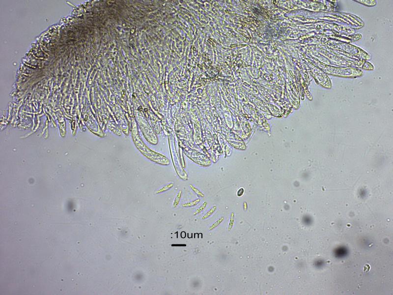
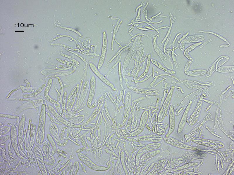
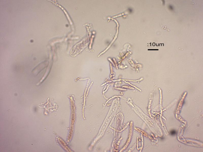
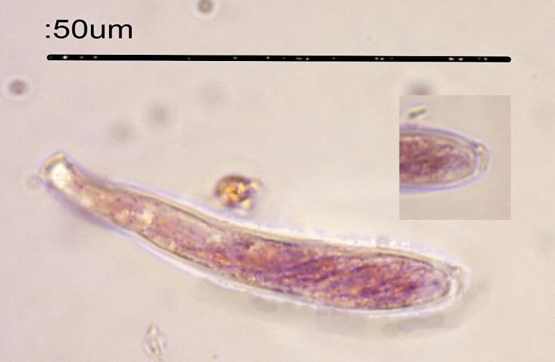
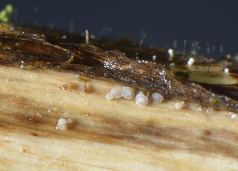
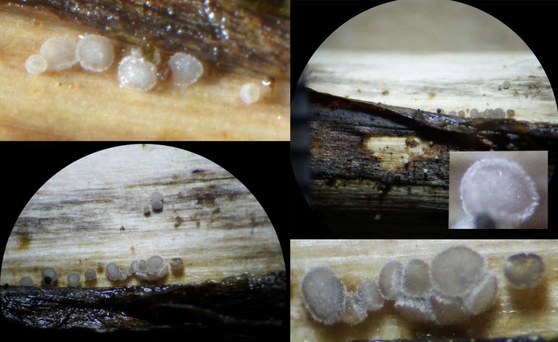
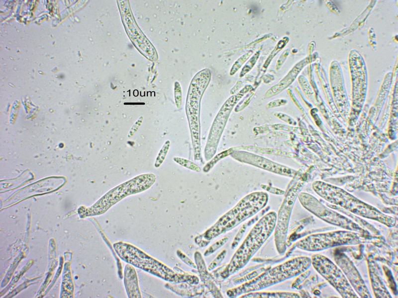
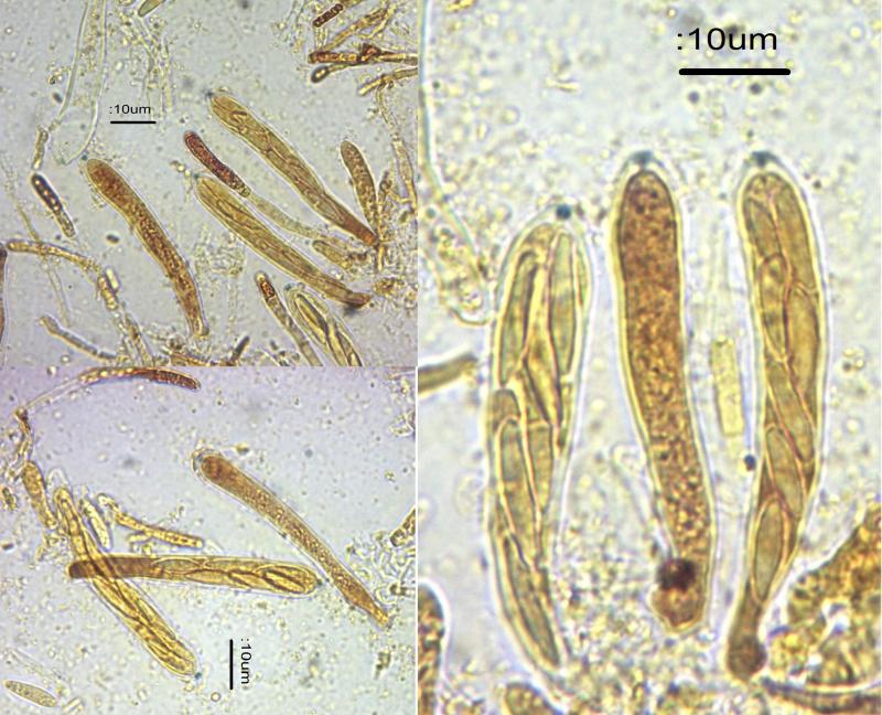
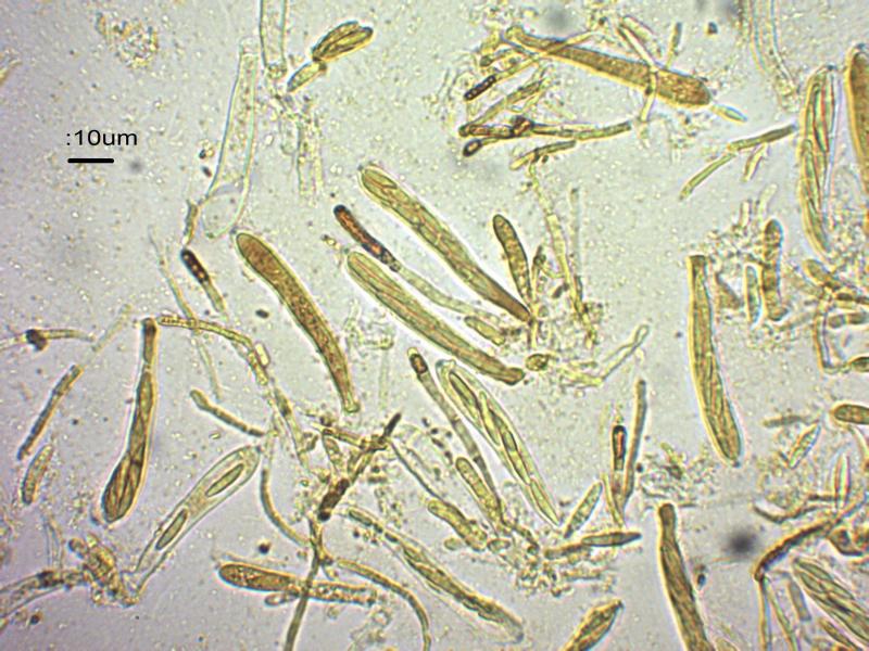
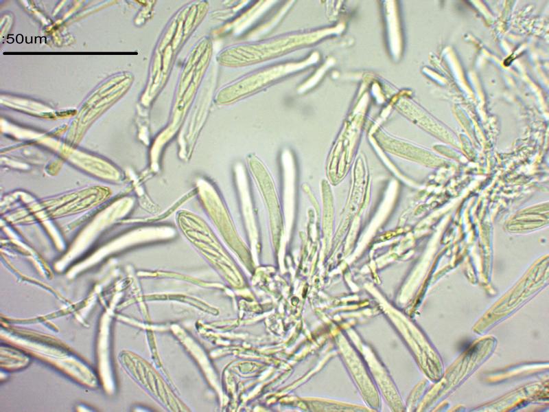
This one has me baffled, so any help would be appreciated. It was on dead stems in a Rosebay Willowherb (Chamerion augustifolium) patch. One small patch of ascocarps, some partly beneath the stem epidermis. Up to 0.3mm (300mu) across. Discs smooth, flattish and pale grey. Margins whitish, granular but not hairy. Some ascocarps clustered. As the fungus was so small I was unable to take sections or samples of parts so I made whole ascocarp squash mounts in water, Lugol and Meltzers. Spores in the squash were 11-16 x 3-4, non-septate. Asci 8-spored, up to 50 x 10 mu, tips bluing in iodine reagents, some look as though they have croziers. Paraphyses appeared tp be cylindrical 2-3 wide, browning in Lugol. There were plenty of branched structures present – paraphyses? From the squash no hairs are apparent.
Ellis and Ellis got me as far as Pezizella, but nothing there has spores which fit.
I got nowhere with Brian Spooner's key from the BMS Asco workshop.
Neither can I find a fit in Peter Thompson's book.
I wonder if this is another Mollisia?
Steve
Hans-Otto Baral,
17-03-2015 23:14

Re : Grey and white Discos on Chamerion augustifolium
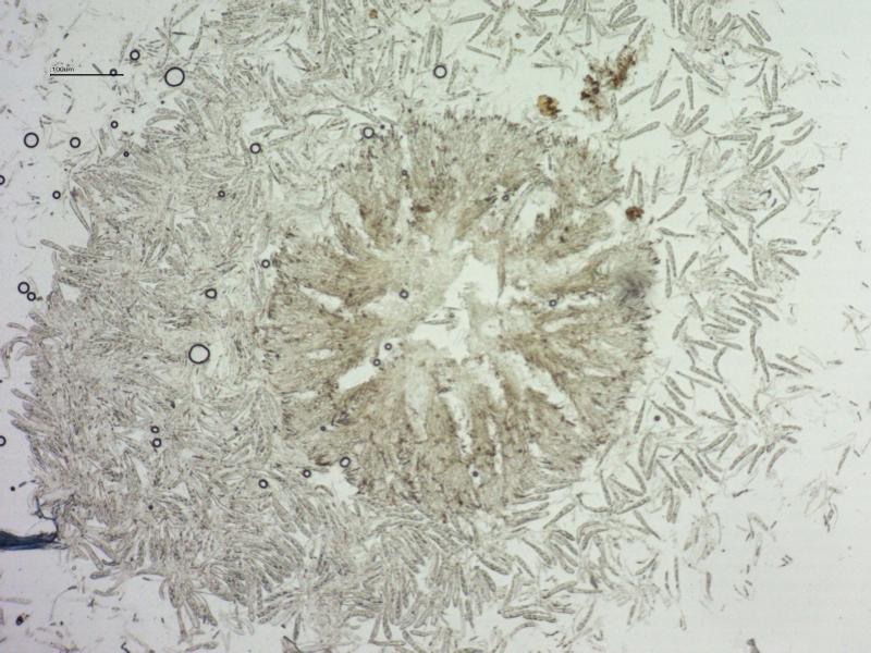
Can yu make a photo of the excipulum in external view? Only a closeup of your survey photo. Also the margin would be necessary if there are any small hairs.
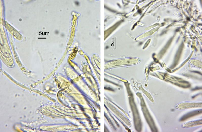
The paraphyses contain distinct VBs (they are what turns red-brown in iodine), therefore it cannot be a Cistella. I compared Psilachnum lanceolato-paraphysatum, but paraphysis shape excludes this, also spore size.
Zotto
The paraphyses contain distinct VBs (they are what turns red-brown in iodine), therefore it cannot be a Cistella. I compared Psilachnum lanceolato-paraphysatum, but paraphysis shape excludes this, also spore size.
Zotto
Steve Clements,
18-03-2015 00:01
Re : Grey and white Discos on Chamerion augustifolium
Thank you, I will examine the excipular cells - it may take a couple of days.
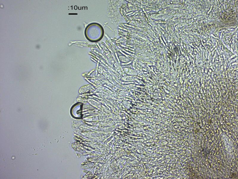
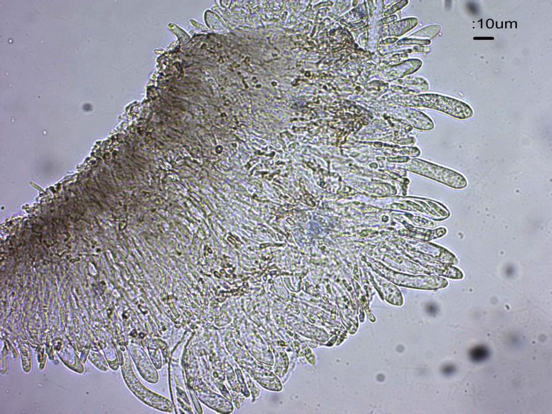
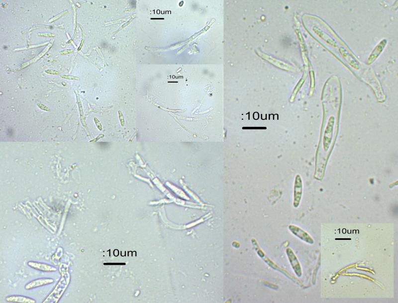
Found some time to look at the last couple of ascocarps. I can't get better pictures than the macrophotos already posted - I used a flashgun, extension rings and additional lens. The ascocarps are very small - mostly less than 2-300 um across. I'm sure that there are no hairs, and that it's the protruding asci at the margins which make them look minutely hairy. Otherwise, all I can see are asci, paraphyses and branched structures. These are images of whole ascocarps, some from the edges, other are squashes.
Cheers,
Steve