

10-06-2026 12:54
Steve ClementsBonjour encore, Pouvez-vous m'aider, s'il vous pl
09-06-2026 18:32
Camille MertensSur morceau de roseau immergé 0,5 - 0,7 mm de dia
10-06-2026 21:16
 François Freléchoux
Bonsoir,Le dernier du jour, en attendant votre avi
10-06-2026 21:07
François Freléchoux
Toutes les tiges de gentianes jaunes de l'an pass�
10-06-2026 13:41
François Freléchoux
Bonjour à nouveau, Voici une trouvaille d'hier.
10-06-2026 11:53
Steve ClementsBonjour, This disco is abundant on dead stems of
10-06-2026 10:45
François Freléchoux
Bonjour à nouveau, Encore une détermination qui
08-06-2026 10:16
 Spooren Marco
I don`t have a clou about this fungus,it is not in
10-06-2026 09:24
François Freléchoux
Bonjour, J'imagine que cette détermination ne do
Hymenoscyphus? with mould on bark
Steve Clements,
19-04-2015 14:10
Hi,
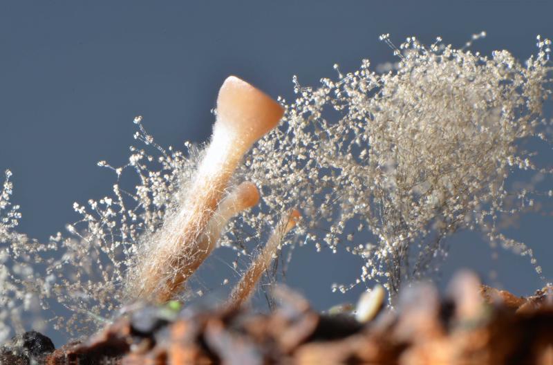
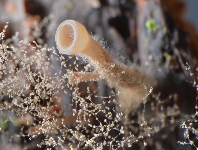
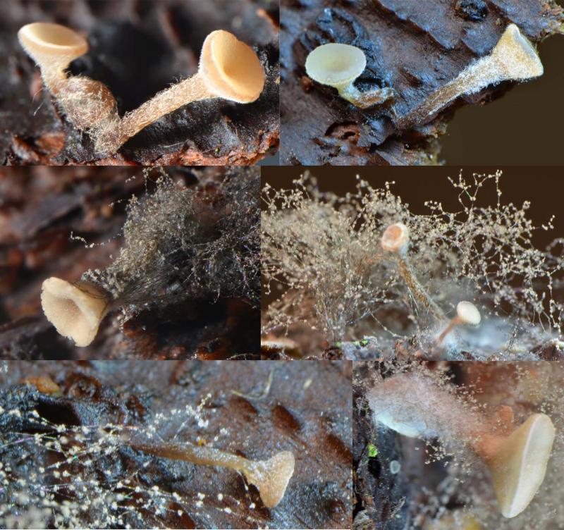
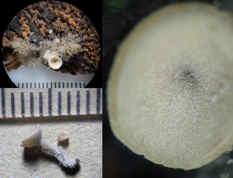
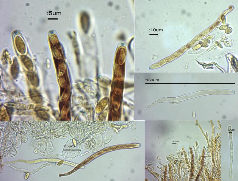
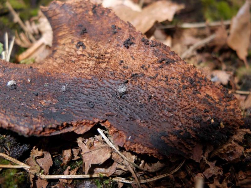
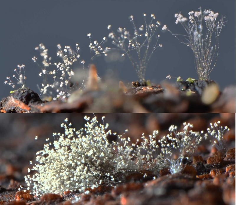
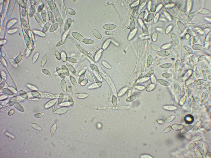
This stalked disco appeared growing with an unidentified grey mould on a piece of bark of unknown provenance (pile of branches), collected for ID of the mould. 5 mould colonies developed, in 4 of them were the stalked discos, developing over 1-4 weeks in an incubator. Up to now 1 mould has no disco, and one pair of discos have developed apart from the mould. The discos are either single of paired at the base. The concave, pallid brown powdery cups are up to 3mm – edges not hairy, with hairy stems up to 5mm long, 1mm wide, browner towards base. Tissue generally soft. Spores are hyaline 11-15 x 5-6 um. Asci seem mostly to be without croziers, up to 150 x 8, tips blueing in Lugol, inoperculate, contents reddening in Lugol. Paraphyses cylindrical, septate, 3-4 wide, rounded ends. A small piece of tissue was carefully removed from the outside of the cup, and showed both narrow hyphae and rounded cells approx 30 x 25 - also noted were some septate terminal hyphae and narrow rounded cells of various sizes. The discos don't appear to be infected with the mould as they remain in healthy growth.
I suspect this is a Hymenoscyphus, but cannot find a species with spores of this size.
Any help would be much appreciated!
With regards,
Steve


Hans-Otto Baral,
19-04-2015 18:00

Re : Hymenoscyphus? with mould on bark
This is sclerotiniaceous. Perhaps the mould is the anamorph, did you try to identify it? Botrytis, for example?
Steve Clements,
20-04-2015 00:05
Re : Hymenoscyphus? with mould on bark
Hi Zotto,
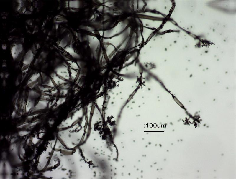
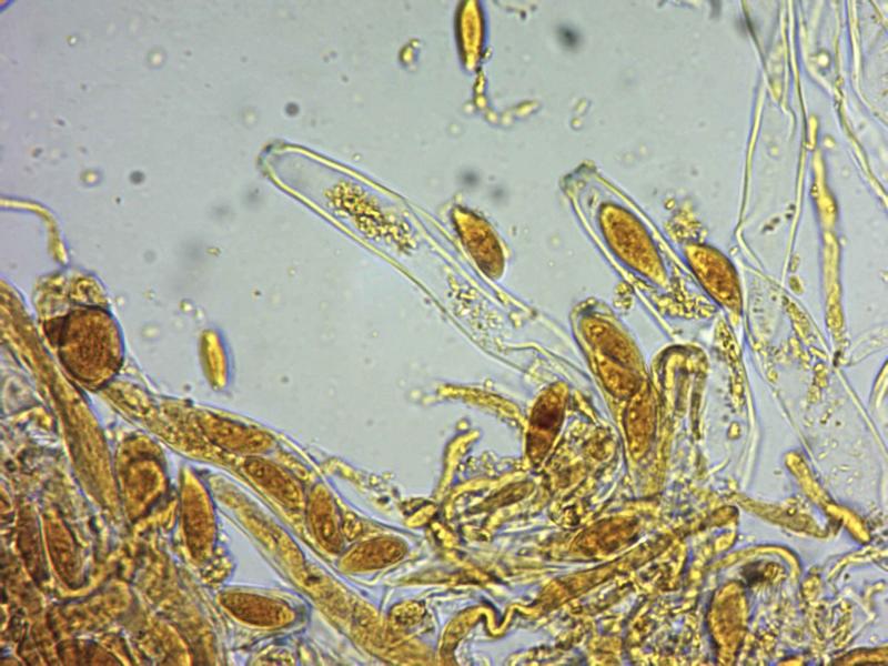
I puzzled over this as it isn't included in Ellis and Ellis as on wood. I think it's Botrytis, and the spores at 8-14 x 7-9 (in water) fit B. cinerea. I was interested in the constictions in some of the hyphae, which aren't evident in my pictures of B. cinerea e.g. Webster (1980). The grey colonies are up to 8mm high, with a tree-like structure, with the "stalks" between 10-20 um across, hyaline (not brown as in B. cinerea as described in Ellis and Ellis). Is "Botrytis cinerea" a collective name for a number of species? Could the stalked disco be Sclerotinia fuckeliana?
Best regards,
Steve

Hans-Otto Baral,
20-04-2015 08:31
Re : Hymenoscyphus? with mould on bark
With constrictions you mean collapsed cells? These are evident on your pics and result from desiccation tolerance and a water-repellent surface, obviously. These hyphae should take up water after a while....
I am not very familiar with Botrytis anamorphs. But it would helb to see living paraphyses, if you have the fungus still fresh. In water mounts made without pressure they should survive. Botryotinia has usually striking contents (VBs). A closeup of the spores (oil immersion=) would also be useful - the spores are possibly 2- or 4-nucleate and nuclei can sometimes be seen, at least when carefully staining with IKI or CRB.
The holomorph name is now Botrytis, but about the species I cannot presently speculate.
The bark looks much like Fagus. You know the place where this pile was?
Zotto
Steve Clements,
20-04-2015 09:25
Re : Hymenoscyphus? with mould on bark
Good morning Zotto,
Yes, the wood pile is in a marshy area in mixed woods at about 1000ft on acidic gritstone rock with peaty soil. Standing trees are mostly Birch and Alder but there are Beech in the vicinity. Today I must look after my granddaughter but I shall do more microscopy later if my material is still in good condition,
Regards,
Steve
Steve Clements,
20-04-2015 22:37
Re : Hymenoscyphus? with mould on bark
Hi Zotto,
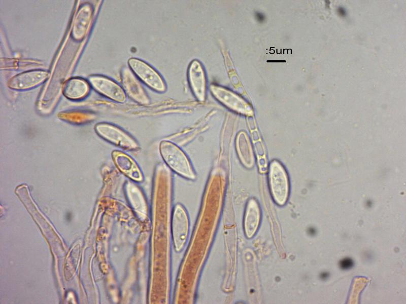
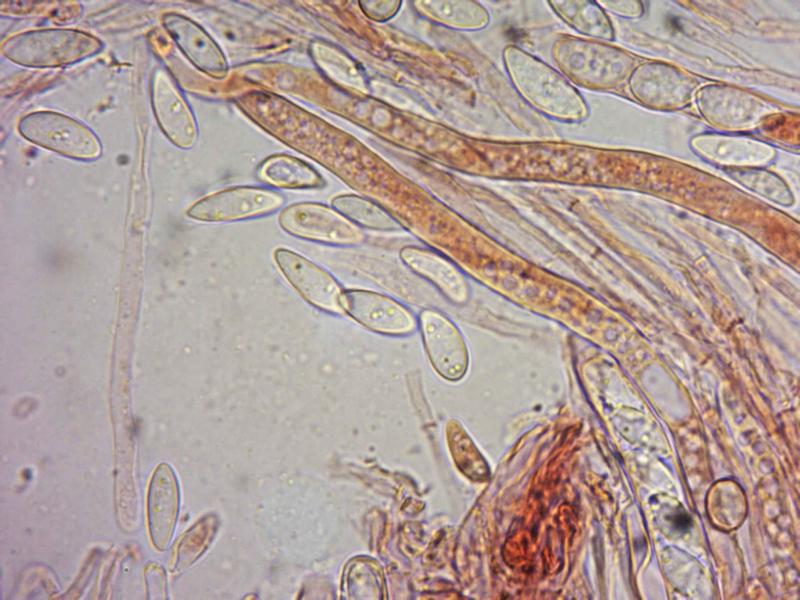
I have used up the last of my ascocarps in making the following pictures. In water the paraphyses are almost invisible - they need Congo Red stain to show up - they are narrow cylindrical, some with slightly swollen heads, about 4 mu across. I've stained the spores in Congo Red and Lugol.
Regards,
Steve


Hans-Otto Baral,
20-04-2015 22:57
Re : Hymenoscyphus? with mould on bark
What I can see is that the oil drops in the spores are sometimes in the middle - and this indicates that at least two nuclei are in the spore.
Living paraphyses are rather well visible without staining. But the feature of VBs gets completely invisible in dead cells....
I have presently no time to go in the literature about this fungus, though I would like to.
Living paraphyses are rather well visible without staining. But the feature of VBs gets completely invisible in dead cells....
I have presently no time to go in the literature about this fungus, though I would like to.
Steve Clements,
20-04-2015 23:20
Re : Hymenoscyphus? with mould on bark
Many thanks,
I shall record this as Botryotinia cf fuckeliana as it is with Botrytis anamorph
Steve
I shall record this as Botryotinia cf fuckeliana as it is with Botrytis anamorph
Steve