

17-05-2026 19:05
Thomas FlammerI have found this tiny 200 ym cup shaped apothecia
17-05-2026 16:41
Margot en Geert VullingsWe found this Lachnum on an old Rubus stem.Fruitbo
05-04-2026 22:46
 Lothar Krieglsteiner
on wood of Ceratonia, Algarve, 3.4.2026.The color
15-05-2026 13:33
Sylvie Le GoffBonjour à tousJe serais très reconnaissante enve
16-03-2011 14:31
roman vargas albertoHi. I would like some opinion about this Peziza
14-05-2026 05:36
Ethan CrensonHi all, I haven't paid much attention to Lachnu
10-05-2026 23:17
 Andreas Gminder
Hello,today we found in a moist steep decidous for
11-05-2026 12:32
 Bernard CLESSE
Pourriez-vous m'aider à identifier cette héloti
13-05-2026 15:26
 François Freléchoux
Bonjour,Voici une récolte faite il y a quelques j
 The study of Moellerodiscus in Malta has so far confirmed M. lentus s.l. on leaves of Ceratonia siliqua L. - both from my own study and that of my friend Carmel Sammut.
Eg:
http://www.ascofrance.com/forum/34675/ciboria-on-ceratonia-leaves
I have now found the Maltese Moellerodiscus on two different hosts, but always leaves:
Olea europaea (olive)
Rhamnus alaternus (Buckthorn)
Maybe these hosts are new for the species and also broadens the knowledge that the species is not host specific (reading that similar M.l. where also found on fruit of carobs)
I also have a question, I have collected the material Saturday but had no time to work upon their micro characters and I shall be abroad for three days. What is the best way to preserve - dry in air or keep moist in a humid chamber?


if you can keep the samples in the fridge at +5-10° they will be o.k. after 3 days. Otherwise I would let them dry and study later. Perhaps you will find living paraphyses or asci after that short time when rehydrating.
Zotto

This species on Rhamnus alaternus I believe it is a different species to the one on Ceratonia leaves. You can see some of my micropictures of it in Zotto´s folder
https://drive.google.com/drive/folders/0B5SeyOEkxxZhQjU0MkMzM2RYNXM
But there is also in that folder another species which grows on Plantago. To distinguish them, read the "descrpt.ion" archive in that folder.
We gained a sequence from the one that grows on Rhamnus alaternus leaves in our area, and it falls shortly apart from the Moellerodiscus on Fraxinus and the one on Hedera (which I believe could match with yours, one of them... or who knows if a different one!). This is also supported by habitat, colour (never yellowish) and a tendence to show more hair-like terminal cells at margin. Besides that, the hemiamyloid reaction of the excipular cells is more evident in this species on Rhamnus.
About the Olea find... I also checked and sequenced a collection on Olea leaves, and it matches perfectly with the one without croziers that grows on Michel´s Malus fruits and our collections on Eucalyptus bark and Ceratonia pods. So check carefully for absence of croziers in that one. Please tell us what the micro says when you have the opportunity. You can maybe let dry some apothecia and keep other in the fridge till you can study them.
Cheers,
Raúl
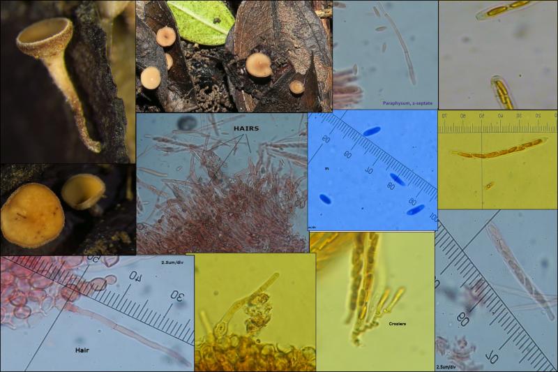
PS: I attach a composition of our Rhamnus collection

[1]
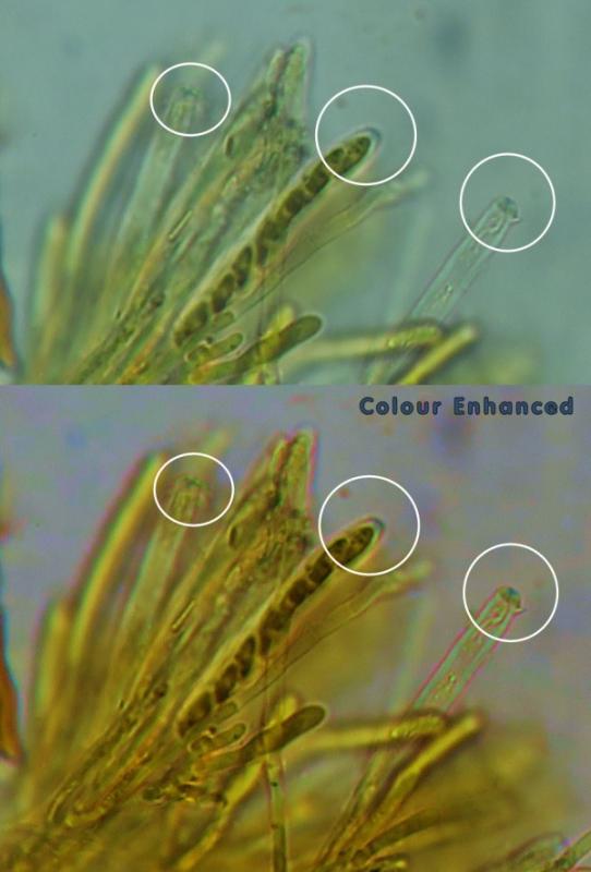
They were rehydrated in water for 30mins which they gained their full vigour and original shape. The IKI test prooved to be faintly positive at the pore. I followed your procedure in another post where the IKI was washed by KOH and water after a second application. This reaction can be easily detected if the colour is enhanced by post-processing (and applying white balance to the background).
[2]
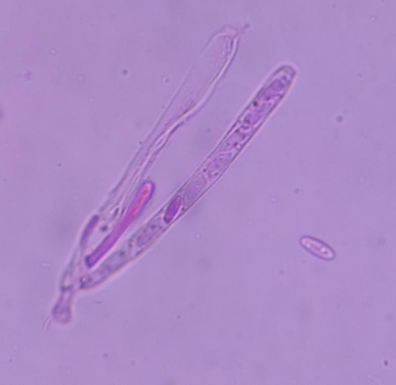
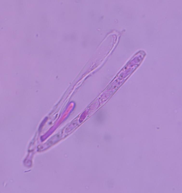
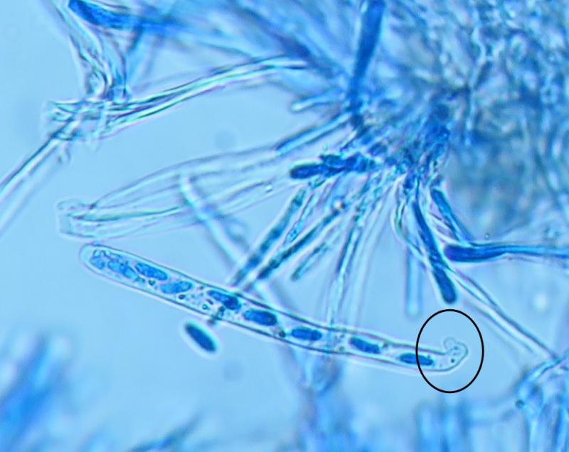
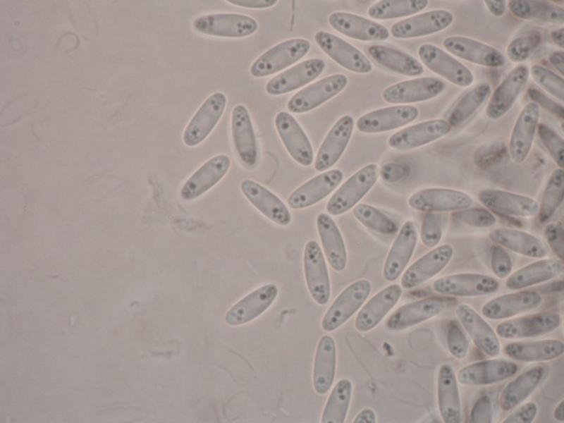
The spores were fusiform, slender, 7-8um long 2-3um wide, rather homogenous or varying slighty in size, with distinct small oil bodies aggregated at the poles. Very hyaline and slightly stained in congo red.
[3]
Asci 62-75 x 5-6um, slightly and gently bent longitudinally.
[4]
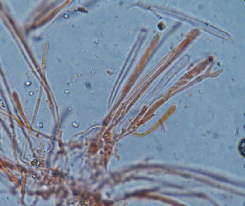
Paraphyses in small bunches, regular width (apex not swollen) and becoming well stained in IKI or cotton blue, less so in Congo red. I think some were septate. Apex undifferntiated.
[5]
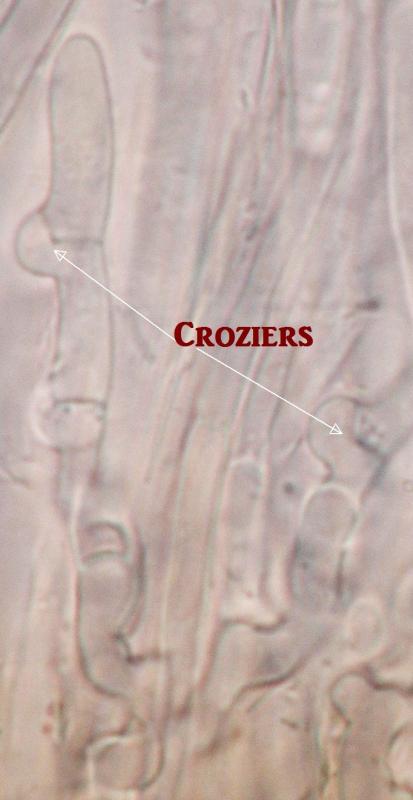
I have not seen corziers (except one doubtful instance) but it is very difficult to find free small-clusted asci to examine their base. I guess highly active croziers would staind very well in cotton blue. When I applied this stain, I only saw jaggered asci bases but no typical croziers.
[6]
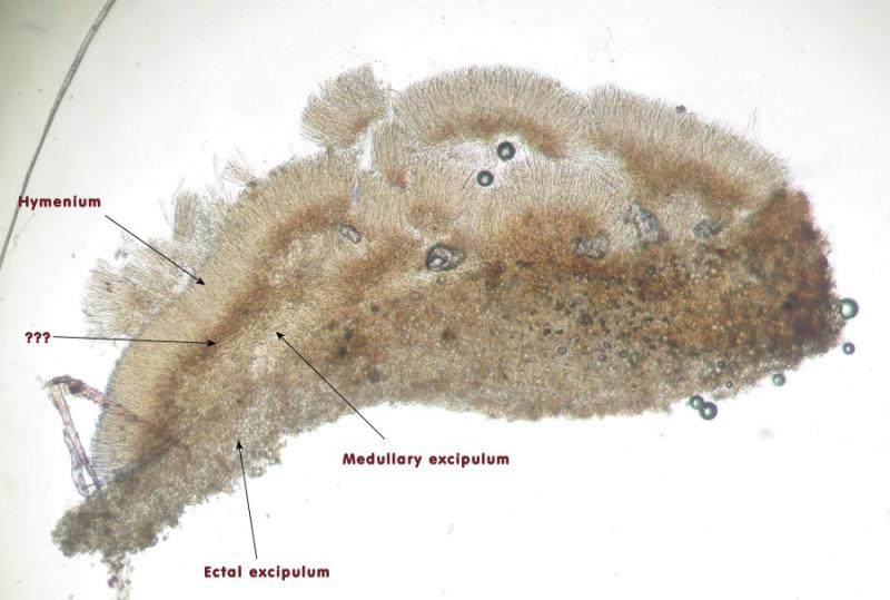
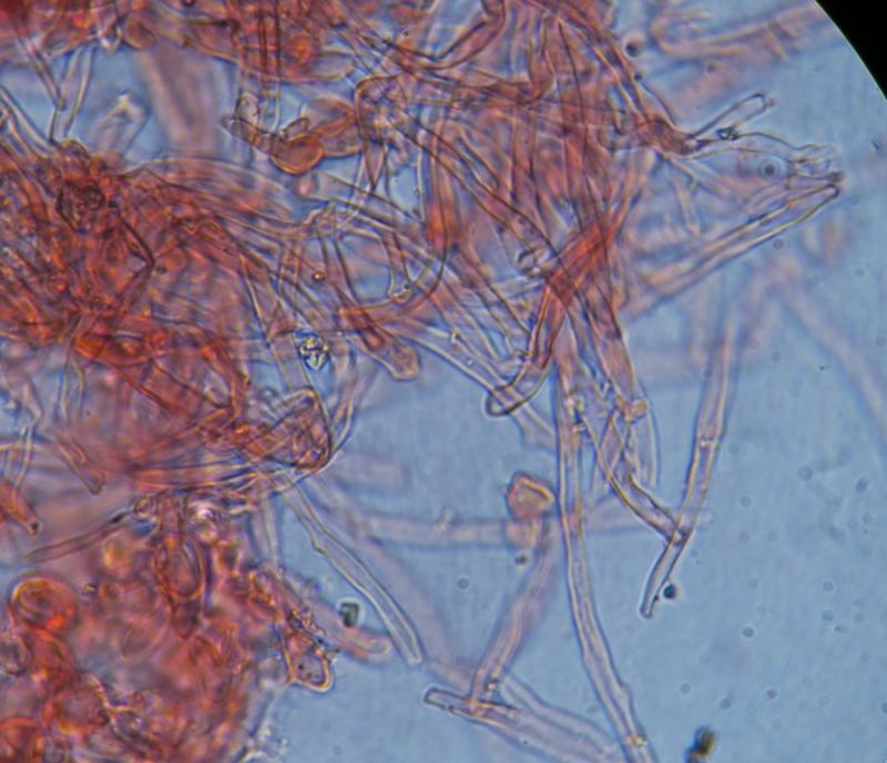
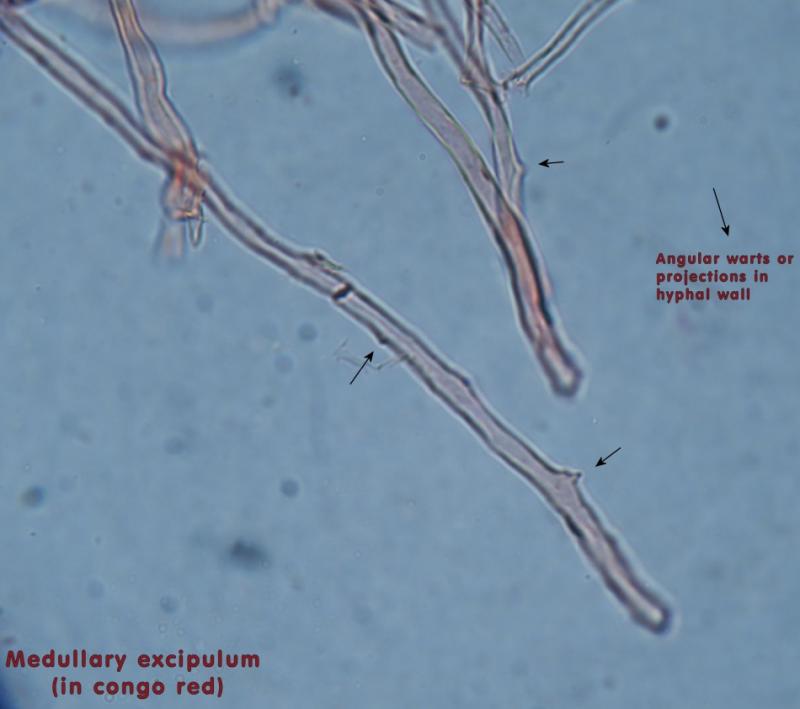
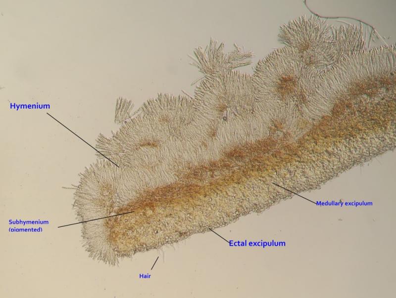
The excipilum consist of two layers:
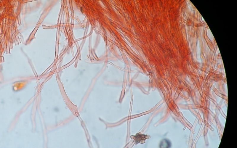
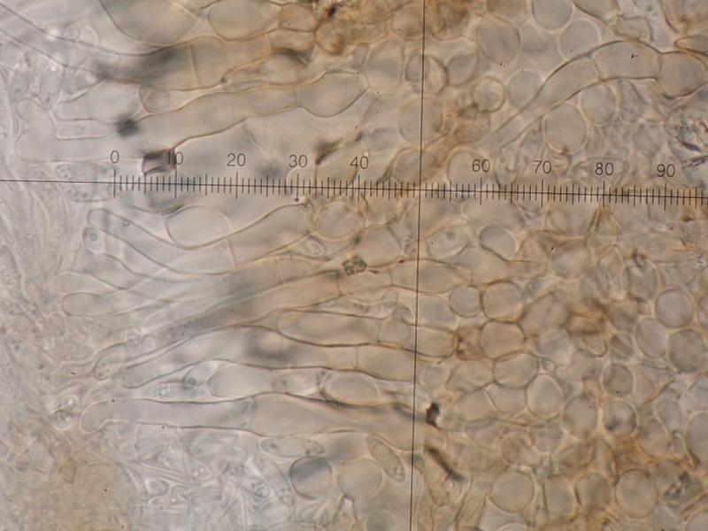
Medullary Excipulum: Spongy thick layer of tessuta intricata. Hyphae intricate irregularly curved or bent tubular hyphae measuring 4-6um in width, with sparse angular warts along their walls (c. 1-4 per cell), inconsistent width and length.
[7]
Ectal Excipulum: Spongy thick layer of tessuta intricata. Hyphae intricate irregularly curved or bent tubular hyphae measuring 4-6um in width, with sparse angular warts along their walls (c. 1-4 per cell), inconsistent width and length.
[8]
When I made a thin cross section across the ascomata, I have noticed a third layer above the medullary excipilum and below the hymenium. Narrow and pigmneted, similar tin structure to the ectal excipulum. What is this layer ?
----------------------------
It seems to be similar to the Moellerodiscus lentus I've recently found on carob leaves. I have judged that the croziers are absent or indistinct. Now I work on the other Moellerodiscus population on Rhamnus leaves.


Thanks
Microscopically, I have not seen much difference, except the hair cells that are longer and the spored marginally longer by about 1um. I don't have the time to upload the photos one by one but I hope the link below works out.
https://drive.google.com/drive/folders/0B_a9JPbpBp5ASm5neTQ4Zkh6eFE?usp=sharing
There are some ocassions were the are bumps at the base of the ascii which give an impression of croziers. Kindly check. So is this always the same M. lentus or there are differences significant enough to delimit varieties, subsp. or even speceis within this complex ???
Thanks for the tip re KOH and Congo... I keep sticking to them in my mycological work.


For this Rhamnus species you should observe a hemiamyloid excipulum, that is, when KOH pretreated the iodine reaction is pale but distinct blue, whereas without KOH it is pale rose-red, but that red reaction is only obtained with Lugol, not with Melzer.
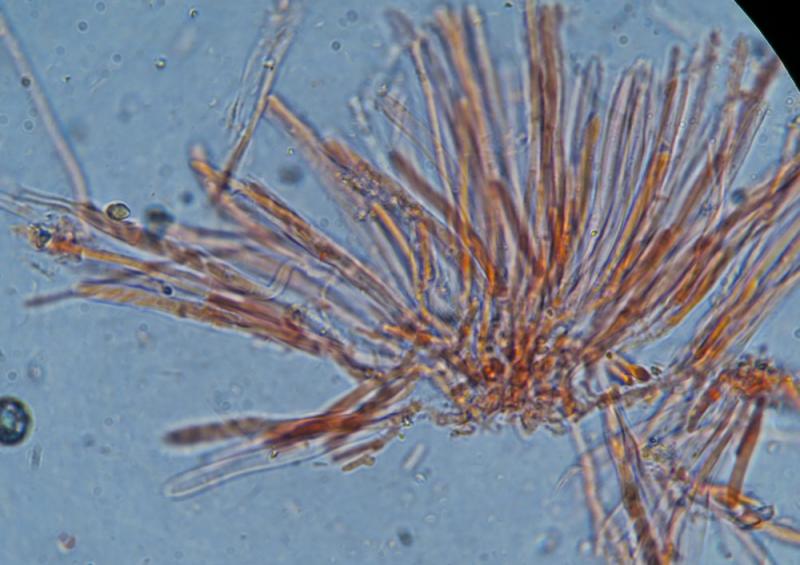
And yes, the hairs seem typical too. I attach some photos made by Raul. Here you can see how the fungus looks in vital state. The pale yellow-brown vacuoles in the paraphyses are characteristic. Note also the sheath around the spores.
Zotto


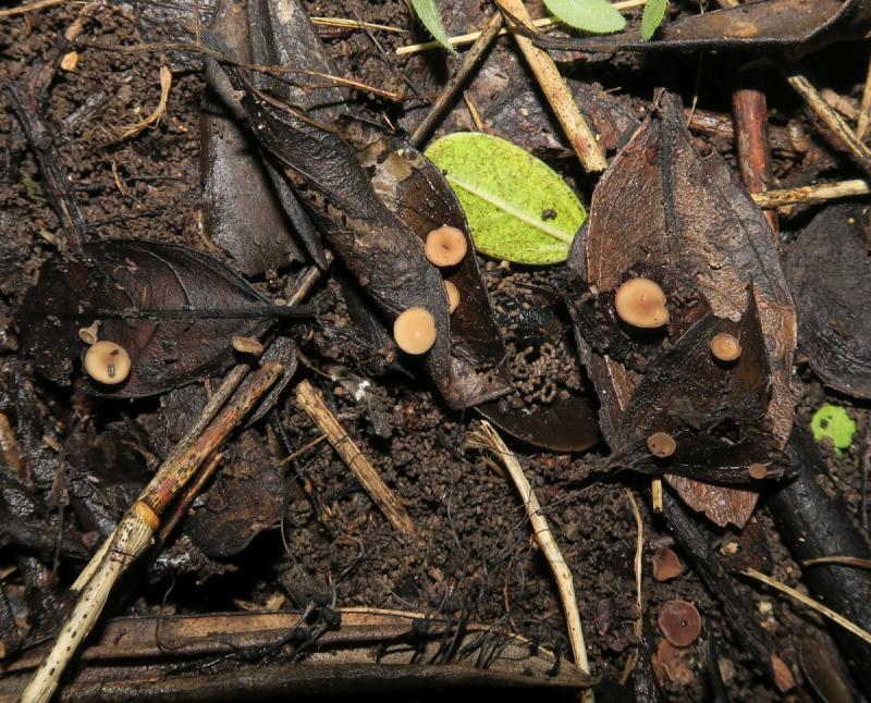
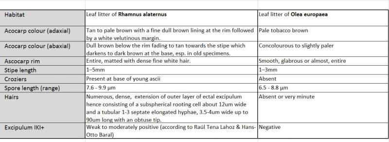
Habitat: Leaf litter of Rhamnus alaternus in a shaded damp area, growing individually on lamina surfaces
Ascocarp type: Apothecium
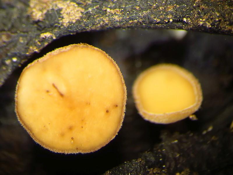
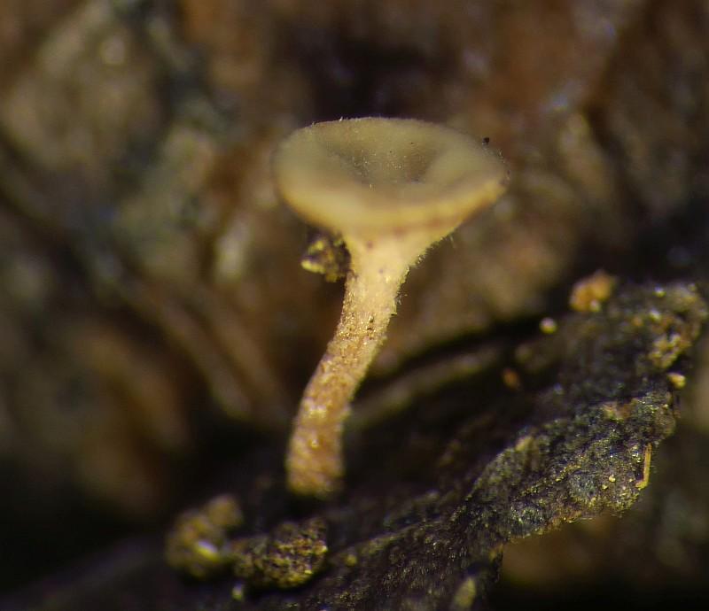
Ascocarp morphology: Stipitate shallow cup to saucer-like ascoma
Acocarp colour (adaxial): Tan to pale brown with a fine dull brown lining at the rim followed by a white velutinous (hairy) margin.
Acocarp colour (abaxial): Dull brown below the rim fading to tan towards the stipe which darkens again to dark brown at the base (esp. in old specimens.)
Ascocarp diameter: 1.56 – 3.81 mm (mean: 2.72 mm)
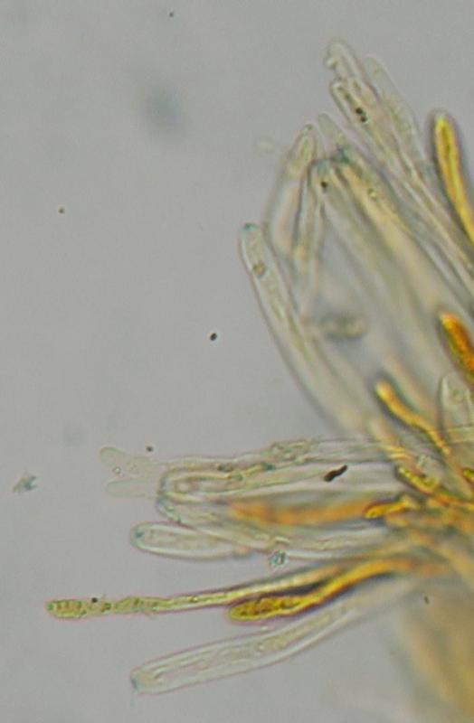
Ascocarp rim: Entire, matted with dense fine white hair.
Ascocarp shape: Shallow cup when young then flattening to a saucer-like shape or just flat. Stipe relatively long and tapering forming an overall funnel-shaped structure.
Stipe length: 1–5mm
Stipe colour: Draken to a medium brown colour towards the bae
Texture: Too small to assess soundly but more or less smooth and coracieous and elastic
Flesh colour: Grey to pale brown
Paraphyses: Numerous, often grouped in small bunches.
Paraphyses shape (apex): Not inflated / differentiated, same thickness throughout its length.
Paraphyses width: 2–3 µm.
Paraphyses length: 70–80 µm, subequal (slightly longer) from asci
Paraphyses septation: Two septa
Croziers: Present at base of young ascii
Ascospore release: Apical orifice without operculum
Shape: Cylindrical, slender, slight bent ot cirved
No. of Spores / ascus 8
Operculum: Absent (inoperculate ascum)
Tunic (Wall): Bi-tunicate
Ascum length (range): 62 - 73 µm
Ascum length (mean): 69 µm
Ascum width (range): 5.7 - 6.3 µm
Ascum width (mean): 6 µm
Ascum L:W ratio: 11.6
Iodine reaction: Weak J+ve only at the apical pore which stains faint blue with Lugol's Iodine after washing with KOH and mounted in water.
Spore length (range) 7.62 - 9.88 µm
Spore length (mean): 8.9 µm
Spore width (range): 2.08 - 3.44 µm
Spore width (mean): 2.8 µm
Spore Q factor (range): 2.51 - 4.26 µm
Spore Q factor (mean): 3.2
Spore shape: Narrow fusiform shape, ocassionally slightly bent laterally
Spore septa: 0 (no speta)
Spore surface: Smooth. Cotton Blue reveals a thick hyaline sheathing wall around spore..
Oil bodies: 2-8, small, oil bodies often gathering at poles of spore. Best visible in Iodine stain, almost undetectable in Congo red
Remarks: Asco carp consists of four layers, a whitish hymenium, sitting on a narrow layer (subhymenium) composed of brown-pigmented hyphae and possily giving the brownish colour of the ascocarp. Below this there is the medullary and ectal excipulum.
Other microscopic observations
Excipulum layers: 2
Medullary Excipulum: Spongy thick layer of tessuta intricata. Hyphae intricate irregularly curved or bent tubular hyphae 3-7um wide, inconsistent variablelength.
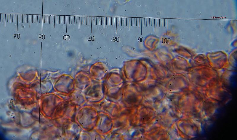
Ectal Excipulum: Narrow layer of tessuta angulata-subsphaerica. Cells pigmented pale brown, isodiametric and angular (but not rounded or spherical), 8-15µm wide (mean 11.8µm), compact, walls roughend with pigment coating.
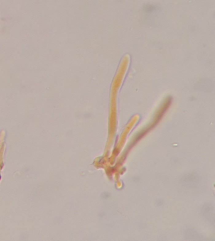
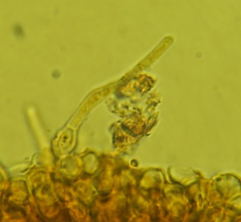
Hairs: Numerous, dense, extension of outer layer of ectal excipulum hence consisting of a subspherical rooting cell about 12um wide and a tubular 1-3 septate elongated hyphae, 3.5-4um wide up to 90um long with an obtuse tip.

Spore length (range) 7.62 - 9.88 µm
Spore length (mean) 8.9 µm
Spore width (range) 2.08 - 3.44 µm
Spore width (mean) 2.8 µm
Spore Q factor (range) 2.51 - 4.26 µm
Spore Q factor (mean) 3.2
Slightly longer from the olea/ceratonia one, maybe not so significant. Does it match with your records ?
Question: What is the pigmented narrow layer below the Hymenium called? I temp. labelled it as subhymenium.
Thanks.
I do not know any Rhamnus collections personally. Also I do not have any project at present about such sclerotiniaceous fungi.
I can take Raul's measurements (5.I.2010 and 28.II.2010):
Asci: 75-100 x 6,5-7,5 or 90-105 x 8-10
Spores: 9-11 x (2,7)3-3,5 or 12-13 x 3-4
The difference in ascus size can be explained by the shrinking effect. be sure that your asci were much larger when alive. Spores do not shrink so much, but it can well be that they were 8-11 x 2.5-3.7 µm when alive.
Zotto
Finally, something curious, I store these ascomata in a closed container. When I open the lid gently, there is some air current/disturbance and this causes the specimes to realeas a small cloud of spores, very visible. Have you experienced this ? I didn't expected this ingenious mechanism from a small and 'simple' fungus, as if they store the ascospores within the ascus and they are only released (with force) with air currents (=> better chance of dispersal).
Thanks
If you happen to find this again, please keep it moist until you study it in oure tap water and without applying pressure.
Thanks for all your images and data! Can you send me two apothecia of both collections? If you don´t mind, I would like to check the absence of croziers in the Olea collection... It looks in some images as it has croziers, but in others not... so it makes me doubt a bit.
Another thing that puzzles me a bit are the colours in your close-ups... they look more yellowish than in the other distant photos. Which are the real ones? In these species I´ve never seen such yellow colours.
About the hemiamyloid reaction... it is hard to see the pink when you add IKI, but the hemiamyod reaction is better seen after KOH-pretreatment. But you have to take care to remove the KOH before you add the IKI again... if not you may do not obtain it because the KOH reacts with the IKI suppressing such reaction. And the reaction is not constant everywhere... sometimes some cells do not react. I find it more constant at the marginal cells.
Cheers,
Raúl
Raúl