

06-04-2026 21:36
 Viktorie Halasu
Hello, could anyone please send me the article wi
06-04-2026 19:40
 David Gibbs
Help with this one much appreciated, on rotting Fa
06-04-2026 11:07
Louis DENYBonjour forum, Trouvé sur bois de feuillu très d
06-04-2026 16:24
Juuso ÄikäsLast Tuesday I found some tiny white Helotiales gr
05-04-2026 20:40
 Robin Isaksson
Hi!Found i Japan on bark of Abies sp. Spores 35-4
06-04-2026 08:15
 Lothar Krieglsteiner
some days ago, on the lower surface of leaf of Que
Mollisia 30-09-18
Blasco Rafael,
06-10-2018 18:53
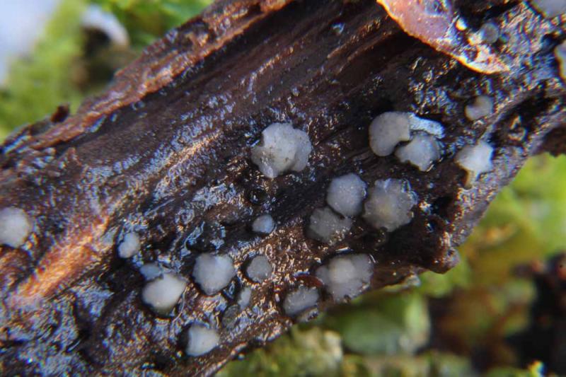
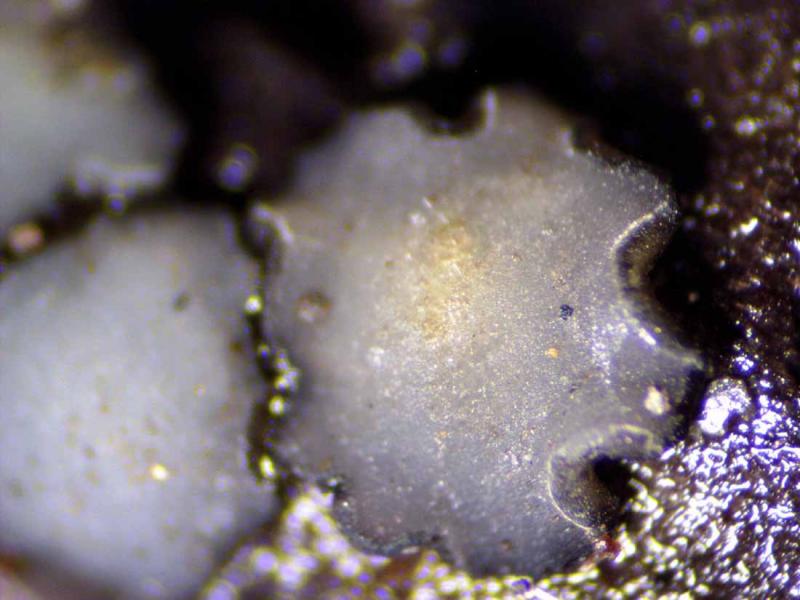
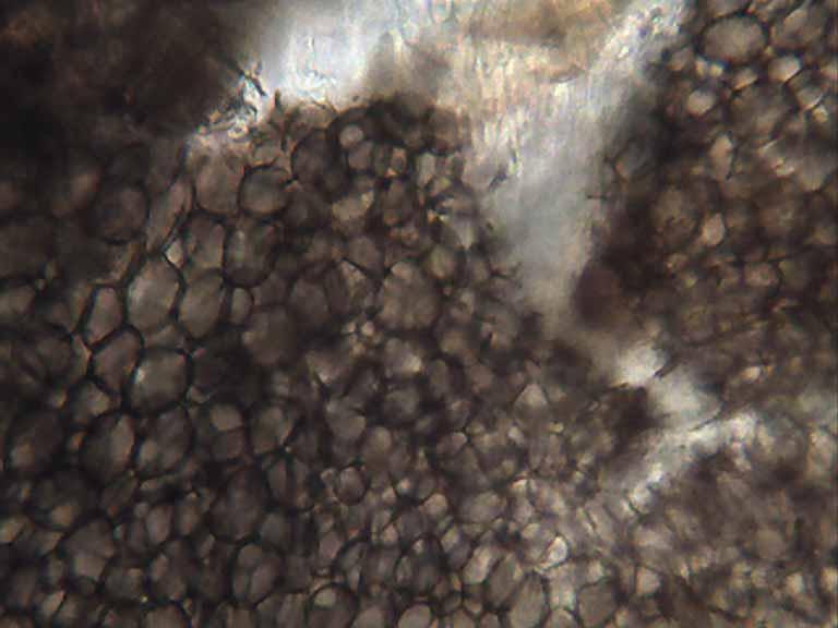
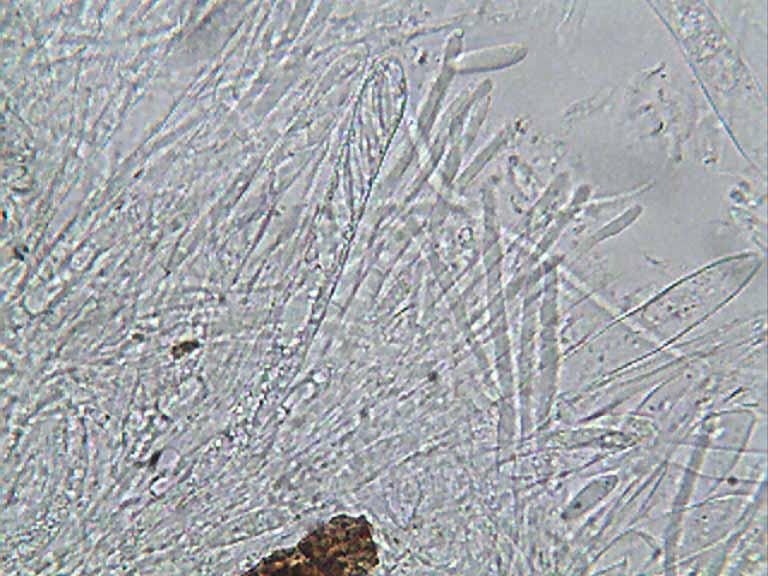
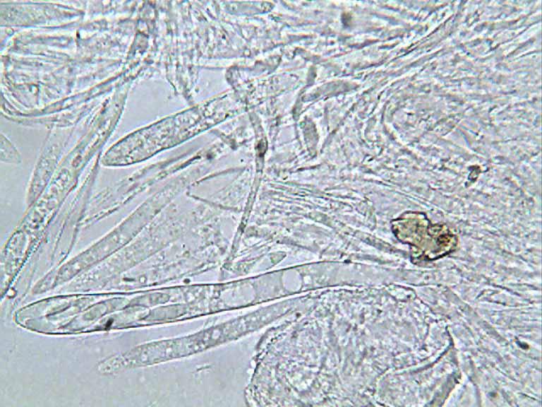
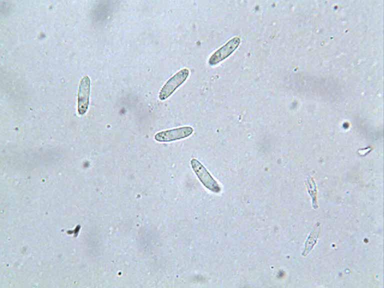
 Hola, alguna idea para esta posible Mollisia??
sobre rama sumergida a 1700m-
diametro hasta 2,5 mm
ascas 90--115 x 10--10,5
esporas 11--15 x 3,9--4,3
parafisis x 3
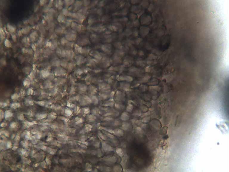
margen y excipulo de celula oscuras de subglobosas a un oco piriformes
Rafael


Hans-Otto Baral,
06-10-2018 20:22

Re : Mollisia 30-09-18
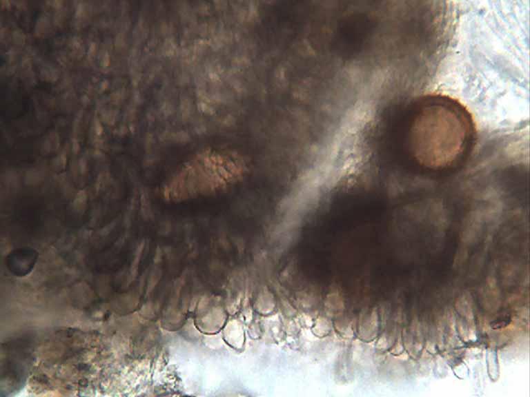
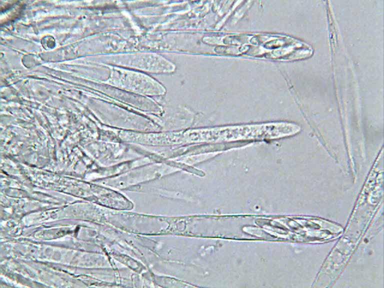
Hmm, I cannot believe such wide spores. But a main probem is: Do the paraphyses contain VBs? I do not see them but there are not enough paraphyses on your pics.
If they contain VBS, and only then you have a Mollisia, you must test the KOH reaction.
If they contain VBS, and only then you have a Mollisia, you must test the KOH reaction.
Blasco Rafael,
06-10-2018 22:14
Re : Mollisia 30-09-18
No veo en ninguna parafisis VBS.
Que otro genero puede ser ??
Rafael
Que otro genero puede ser ??
Rafael
Hans-Otto Baral,
06-10-2018 22:58
Re : Mollisia 30-09-18
That is the good question. Pyrenopeziza on wood, a rare case....
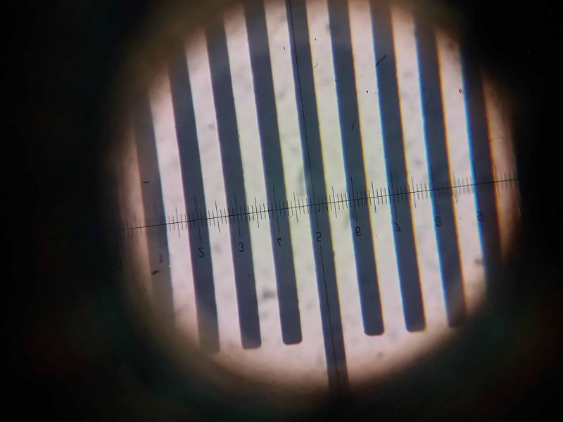
Your micros always have the same magnification, did yi ever ask you for a scale for them in order to remeasure the spores?
Your micros always have the same magnification, did yi ever ask you for a scale for them in order to remeasure the spores?
Blasco Rafael,
06-10-2018 23:28
Re : Mollisia 30-09-18
Hola Zotto
las fotos su siempre X100 en inmersión de aceite
La escala es un tema que tengo pendiente
Rafael
las fotos su siempre X100 en inmersión de aceite
La escala es un tema que tengo pendiente
Rafael
Hans-Otto Baral,
07-10-2018 07:53
Re : Mollisia 30-09-18
If you have the opportunity to use a calibration slide you simply need to take a photo under oil immersion (100x, with oil).
A sample at high altitude with a similar spore size was presented by Yannick Mourgues:
http://www.ascofrance.com/search_forum/11995
But here the spores were septate.
A sample at high altitude with a similar spore size was presented by Yannick Mourgues:
http://www.ascofrance.com/search_forum/11995
But here the spores were septate.
Blasco Rafael,
07-10-2018 10:22
Re : Mollisia 30-09-18
Parece que en 100 micras, falla 2,5 micras
Rafael
Rafael
Hans-Otto Baral,
07-10-2018 10:35
Re : Mollisia 30-09-18
o.k., but
1. The thick scale is in your ocular. That scale is not on your pics, so you take your photos through another ocular? Calibration must be done with exactly the same lens combination.
2. You must send me the spore photo of your "Nollisia" along with the scale photo, because ASCOFRANCE reduces the size of the pics at different rates.
Blasco Rafael,
07-10-2018 11:13
Re : Mollisia 30-09-18
La foto es del Movil, esta realizada para ver que esta bien calibrada.
la lente de calibracion esta en un ocular y la camara en el tubo vertical, con la camara no me sale la medicion,
tengo que retomar lo de colocar escala en las fotos.
como digo siempre estan realizadaas x 100, y periodicamente comprobado que esta escala no varia
Rafael
la lente de calibracion esta en un ocular y la camara en el tubo vertical, con la camara no me sale la medicion,
tengo que retomar lo de colocar escala en las fotos.
como digo siempre estan realizadaas x 100, y periodicamente comprobado que esta escala no varia
Rafael
Hans-Otto Baral,
07-10-2018 11:23
Re : Mollisia 30-09-18
o.k., but you must know that measurements from the ocular micrometer are sometimes different between people, and the only way that I can control the measurement if a scale is included in the photo. If you send me a photo of the scale made with your camera in the vertical tube along with a spore photo of that fungus, I will put the scale in the photo and then measure te spores with a ruler.