
03-06-2026 14:39
Thomas FlammerApothecia yellow, glassy-transparent, 80 - 120 ymS
16-03-2014 13:39
 Enrique Rubio
HI to all I'm looking for B. Hein's article on Wi

02-06-2026 14:33
 Nicolas VAN VOOREN
Hello.I'm searching for a PDF copy of the followin
18-10-2022 00:12
Valencia Lopez Francisco JavierHola amigos/asRecientemente encontrûˋ esta colecci
02-06-2026 17:58
Louis DENYBonjour forum, Sur feuille de Populus tremula, en

28-07-2011 18:31
 Alex Akulov
Dear FriendsToday I made the pdf file of Velenovsk
 Good afternoon.
Does anyone know this anamorph?
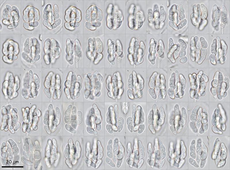
It grew on very humid wood of Erica arborea in Madeira (Portugal). At first I thought it was a Vibrissea.
The conidiospores are together in groups of 6-8, with 1-3 septa. These groups remind me of the fruit of a nut. Over time they continue united, they do not separate, although there does not seem to be an envelope that keeps them together, only hyphae around them. They do not react to IKI, they are yellow.
Thanks in advance.


Let's see if someone is encouraged with new measurement data and some new photos.
The group of conidiospores is composed of 4 long cells with 3 septa and 4 short cells with 1 septum, the measurement of the group is (22.9) 24.2 - 28.8 (30.8) û (14) 15 - 17.5 (18.7) ôçm; Q = (1.4) 1.44 - 1.8 (2) ; N = 62; Me = 26.7 û 16.3 ôçm ; Qe = 1.6
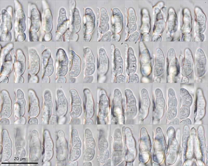
The measurements of the long cells are (13) 14.2 - 18.9 (22.2) û (4.7) 4.9 - 6.3 (7) ôçm; Q = (1.9) 2.6 - 3.6 (4.4) ; N = 63; Me = 16.3 û 5.5 ôçm ; Qe = 3
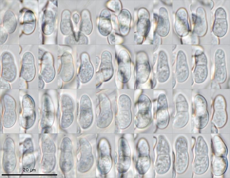
The measurements of the short cells are (8.1) 9.9 - 13 (14.9) û (4.3) 4.6 - 5.4 (5.8) ôçm; Q = (1.7) 2 - 2.7 (3) ; N = 48; Me = 11.7 û 5 ôçm ; Qe = 2.4
I have the impression that at some point in development both parts are joined and then separate into the long and short units.
Any clues?
Miguel ûngel Ribes
cheers
I will check that.
Miguel ûngel Ribes
I think you got the right genus. The type of development and the formation of the conidia, I think, leaves no doubt that it is the genus Amallospora, created in 1897, or something very close.
The curious thing is that this genus only has one species, A. dacrydion, but with a set of conidia that is much larger, 50-75 microns, while mine are 27 x 16 microns.
With this position in the classification (Incertae sedis, Incertae sedis, Incertae sedis, Incertae sedis, Pezizomycotina, Ascomycota, Fungi) it is difficult to look for related genera.
Thanks a lot.
Miguel ûngel Ribes.