
27-04-2026 20:52
 Lothar Krieglsteiner
Found on hanging tiwg of Olea europaea in dried-ou
27-04-2026 18:48
Tony MoverleyCollected 23rd April 2026, Norfolk, EnglandSwarms
27-04-2026 17:41
Lothar Krieglsteiner
.. Algarve, same leaf than the last post. The con
27-04-2026 18:05
Lothar Krieglsteiner
... still attached at standing tree. The green con
27-04-2026 17:16
Lothar Krieglsteiner
.. Algarve, moist lying.The conidiomata look like
27-04-2026 12:54
Steve ClementsBonjour. Ce petit champignon blanc résupiné et
27-04-2026 09:59
Pauline. PennaBonjour Can anyone advise me on these pycnidia fo

22-04-2026 20:54
 Enrique Rubio
Hi to everybody.This Pyrenopeziza grew in moist le
hemiamyloid Chlorociboria from Norway on Picea
Lothar Krieglsteiner,
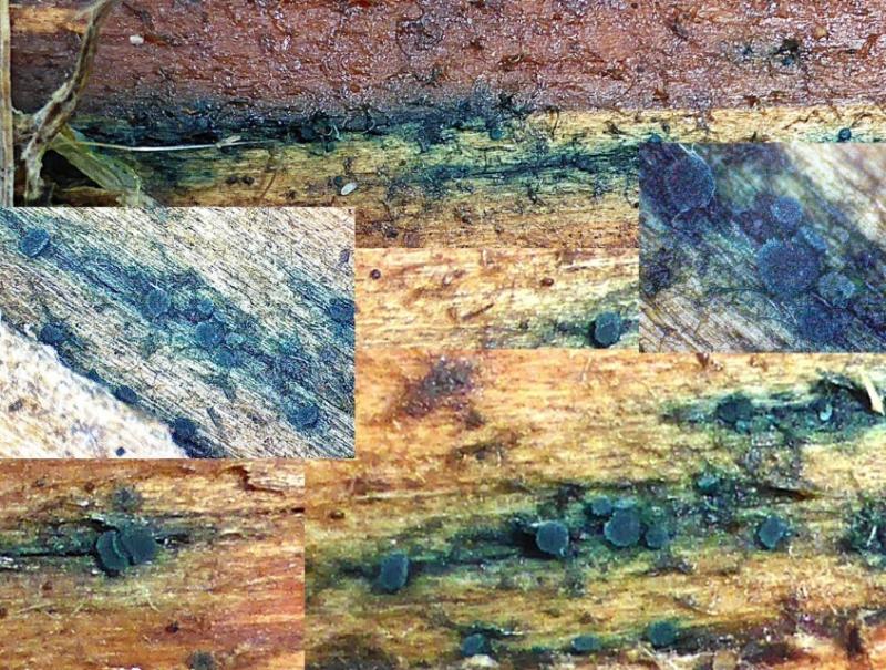
18-08-2025 15:35
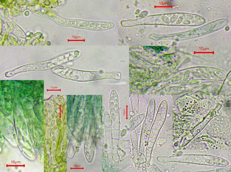
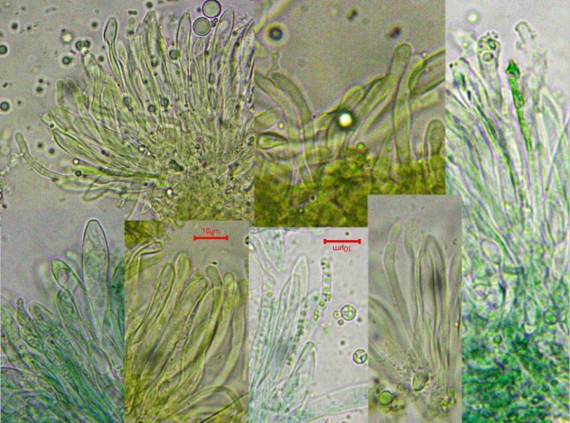
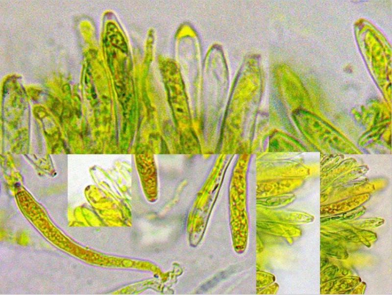
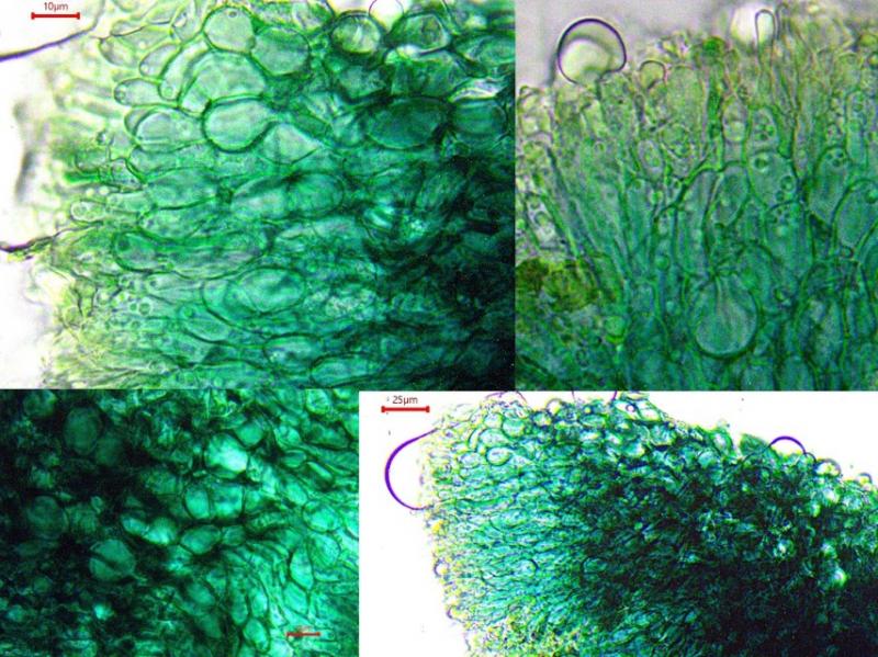
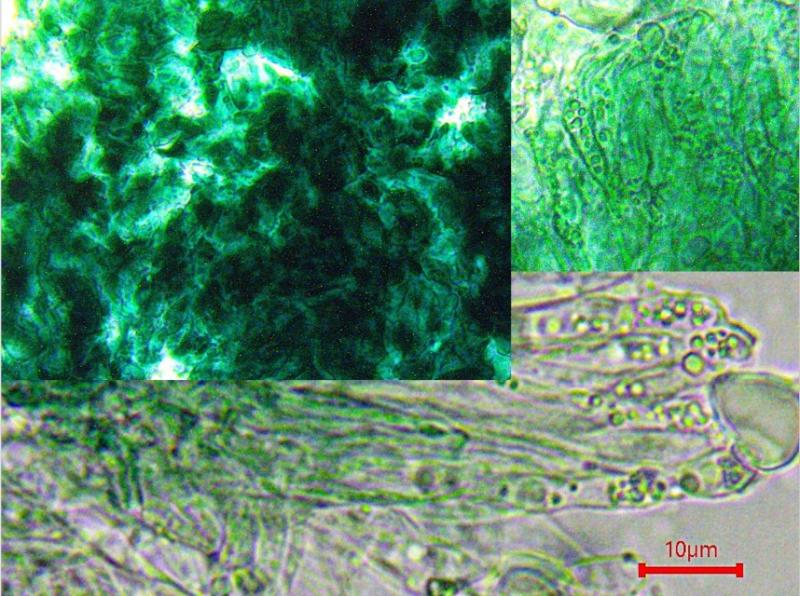
.. in subarctic forest at side of small stream, acid conditions. The asci are brightly hemiamyloid and bear croziers. The excipulum is of blue-green (likely Xylindein as in other species) textura globulosa, in KOH the color is fading to unspectacular olive-greenish. There is no (!) ionomidotic reaction (see below).
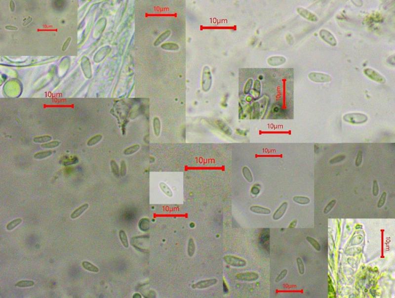
The spores contain few small drops in living stage, in dead stage they are empty. I measured (hopefully not yet shrinked, see below) a size of 5-8(9)/(2)2,5-3,2 µm.
Paraphyses and excipular cells contain some guttules in living stage, they vainsh in dead stage.
I had some problems with this quite scanty specimen. In the first two slides nearly all asci ejected their spores quite rapidly. The free swimming spores would not be calm and could not be photographed satisfactorily. Otherwise the excipular structure contained a lot of exsudate so they could first not be observed well. But in the end I have got most details. The spore fotos are mostly dead spores.
In this forum I found a similar contribution:
http://www.ascofrance.fr/search_forum/72918#
The apothecia found by Andreas Gminder are larger, (mine were up to about 1 mm large) and I found no ionomidotic reaction. In my collection the porus is reacting beautifully red, Andreas noted dirty red. In other respects both collections could be the same species, both on Picea in cold habitats.
Does somebody have a species name here - or is this to be considered undescribed?
Yours, Lothar
Hans-Otto Baral,
18-08-2025 21:09

Re : hemiamyloid Chlorociboria from Norway on Picea
Hallo Lothar
I have no idea but I think the guttules (I assume VBs) in paraphyses and marginal cells are important and would be extraordinary in Chlorociboria. Anyway that genus I would not rule out completely. The VBs remind me of "Cudoniella" buissonii, an otherwise ver different fungus.
I assume it is a fallen hygric branch.
Zotto
Lothar Krieglsteiner,
19-08-2025 08:48
Re : hemiamyloid Chlorociboria from Norway on Picea
Hello Zotto,
thank you very much for your optinion.
"Cudoniella buissonii" (viridula) I once had found - I agree it is quite different at least habitually.
The branch or maybe mor likely root part was moist and it stuck in the earth of the stream side where I had to break it from. First reason was a corticioid fungus, I saw the green wood and the disco only afterwards.
So another not determinable specimen - ...
Yours, Lothar