
11-01-2022 16:36
 Jason Karakehian
Hi does anyone have a digital copy of Raitviir A (
20-05-2026 17:47
Margot en Geert VullingsWe found this Mollisia on dead Juncus stems mown l
22-05-2026 14:47
Gernot FriebesHi,superficial ascomata collected on bark of a liv
22-05-2026 14:44
 Lothar Krieglsteiner
in unripe condition citrine yellow, then soon fadi
22-05-2026 13:29
Gernot FriebesHi,I am curious to hear your opinion on this mater
22-05-2026 10:59
 Nicolas VAN VOOREN
Trouvé sur Phragmites, ce que je pense être un L
20-05-2026 21:49
Margot en Geert VullingsWe found this Lachnum on Juncus stems mown last ye
21-05-2026 17:01
Pierre RepellinBonjour à toutes et à tous,Je recherche l'articl
20-05-2026 20:08
 Andreas Millinger
Good evening,another quite distinctive find from M
20-05-2026 12:57
 Ingo Ibelshäuser
Hello everybody, on decayed hardwood e.g. Quercus
Claussenomyces sp. with reddish brown pigments
Ethan Crenson,
21-10-2018 23:44
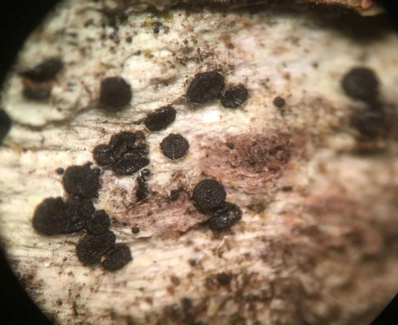
Hello all,
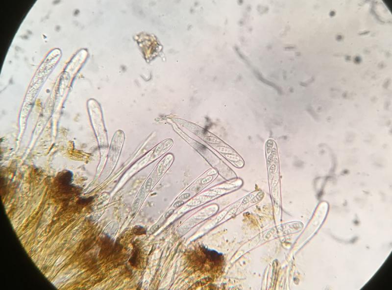
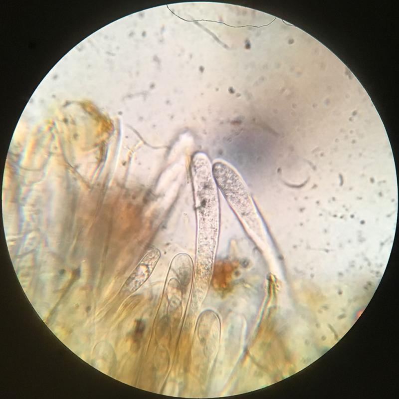
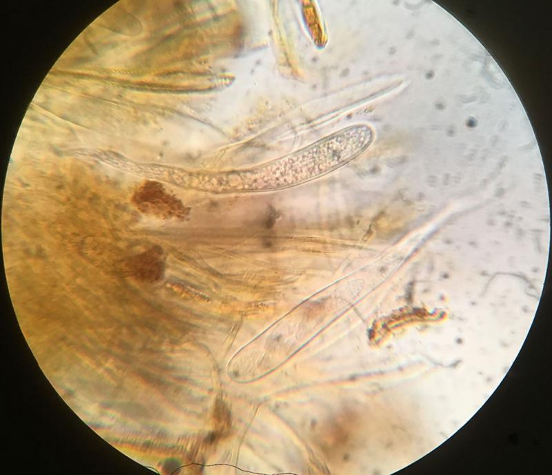
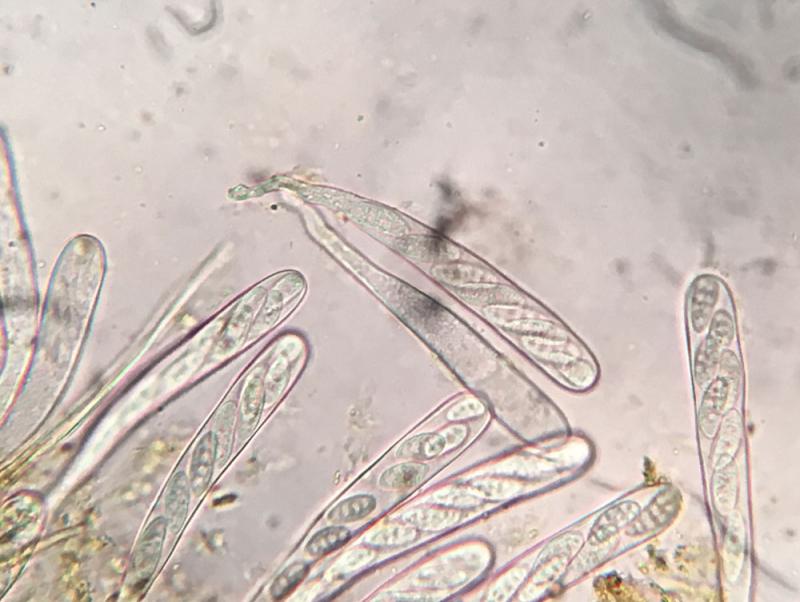
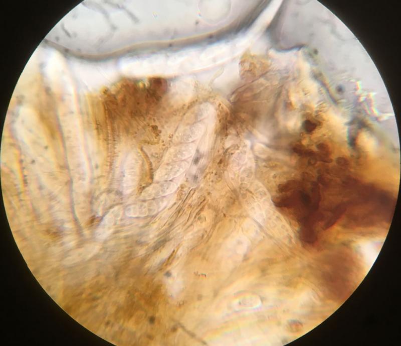
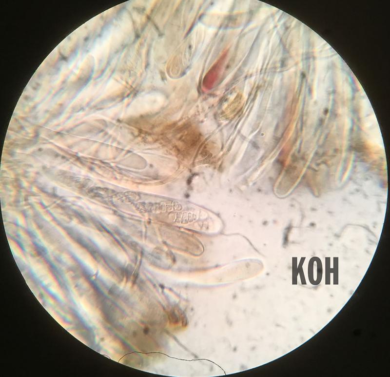
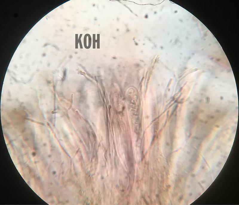
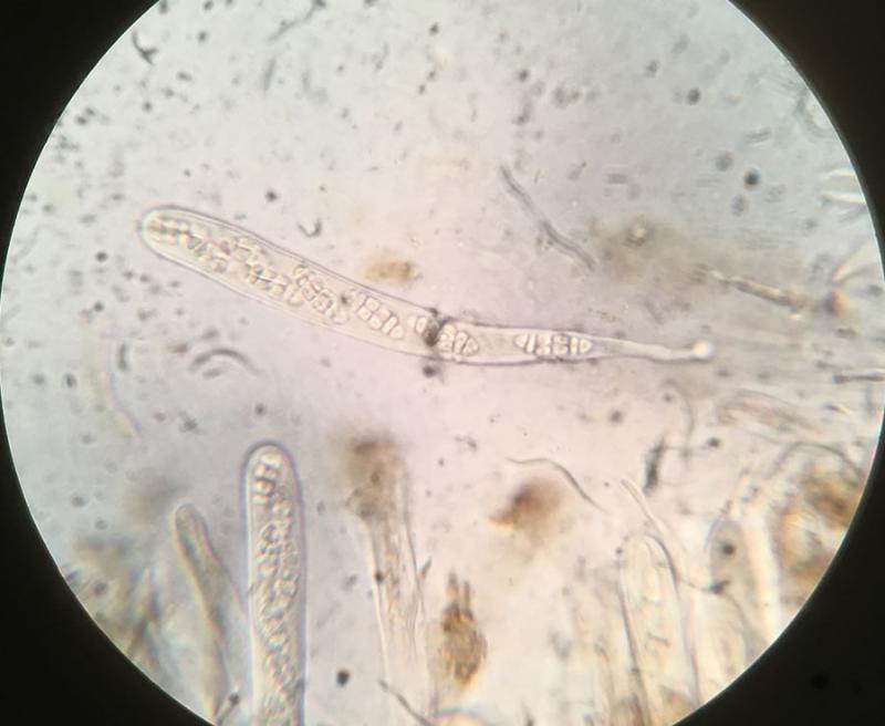
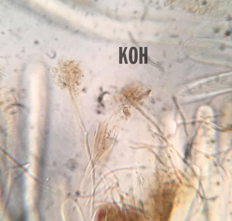
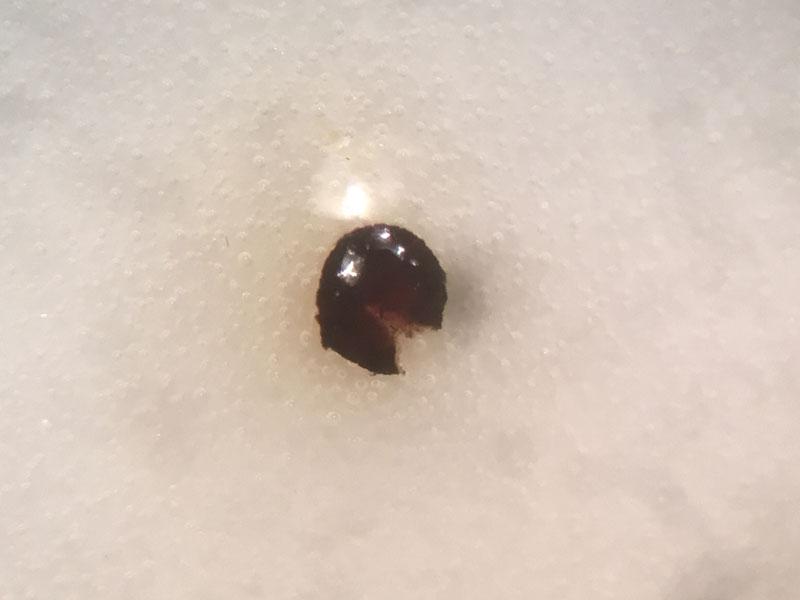
Yesterday in Eastern New Jersey (US), I found a number of very small black discs on decorticated wood. The are about .5 mm in diameter, some smaller, one approaches 1mm. Microscopically they remind me of Classenomyces, with spores that appear at first fusoid and single septate. They then develop multiple septations (5-6 septate) with vertical septations in the center cells. Finally they appear to break apart into tiny (perhaps globose, hard to see) part spores. Asci are around 100-136 by 12-13 µm. Ascospores—the most mature multi-septate ones—measured in ascus were around 16-21 by 7-8 µm. Paraphyses were surrounded by brownish red material. Application of KOH appeared to dissolve most of it.
I tried the key "Provisionary key to Claussenomyces s. auct. (G. Marson + H.O. Baral 17. März, 1994, improved 14.09.1996 and 2002)" which suggested C. canariensis ... but perhaps I am not using it correctly.
Any ideas on this?
Thank you in advance.
Hans-Otto Baral,
22-10-2018 09:13

Re : Claussenomyces sp. with reddish brown pigments
Hi Ethan
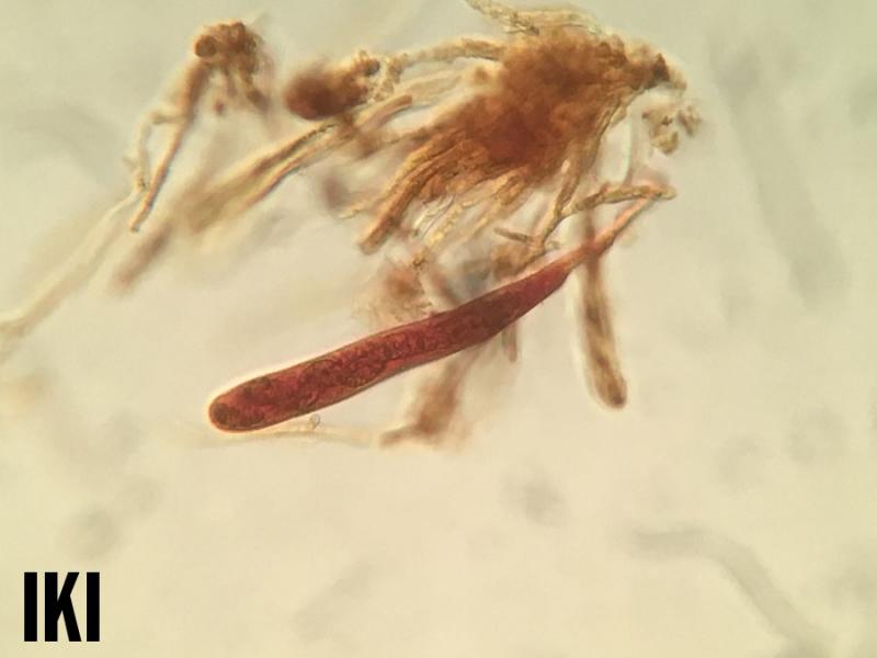
this is an interesting fungus. I assume that the asci are inamyloid in Lugol (did you test it?). Also the dissolution of the brown pigment probably causes a short-lasting brown stin to the medium (ionomidotic).
It could be a member of Cordieritidaceae, and here in the genus Skyttea, but I do not remember muriform spores in this family. Clearly the spores of your fungus get muriform within the living asci.
But there are other families with the same KOH reaction. I think our Claussenomyces-key, in which the ionomidotic reaction rarely occurs, is not the right place to identify this fungus.
Zotto
Ethan Crenson,
23-10-2018 17:14
Re : Claussenomyces sp. with reddish brown pigments
Zotto,
It is inamyloid in Lugol's solution. I tried applying KOH directly to the fruiting body to attempt to see the ionomidotic reaction, but unfortunately I got very little pigment, a very faint reaction if any. Perhaps my interpretation of the previous mount was in error.
Ethan
Hans-Otto Baral,
23-10-2018 17:37
Re : Claussenomyces sp. with reddish brown pigments
If you looked immediately after placing the apo in KOH then it is actually weak. Another possibility is to add KOH to the water preparation and take a pohto in the first seconds when the KOH reaches the tissue.