
28-07-2011 18:31
 Alex Akulov
Dear FriendsToday I made the pdf file of Velenovsk
25-12-2019 17:54
Valencia Lopez Francisco JavierHola a todos/asEstas supuestas pezizas estaban en
12-07-2015 00:05
 Nedim Jukic
This one from the same locality as the previous on
12-11-2019 10:32
 Miguel Ángel Ribes
Hi againExactly at the same place than my previous


30-05-2026 21:12
Philippe PELLICIERSur branche de mélèze (Larix) près de la neige,
31-05-2026 10:35
Hulda Caroline HolteHello,I collected this species growing on a rather
25-05-2026 16:35
 Bernard CLESSE
Bonjour à toutes et tous,J'ai trouvé récemment,
29-05-2026 15:35
daniel FERREBonjour à tous,Je voudrais votre aide pour cette
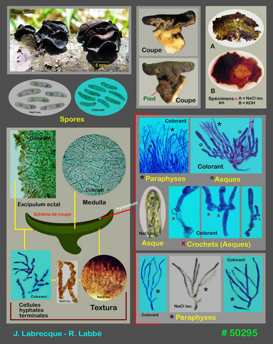
50295 - Ionomidotis fulvotingens ?
Roland Labbé,
27-05-2010 19:43
Voici une planche d'un Ionomidotis.
C'est peut-être I. fulvotingens.
Existe-t-il des espèces dans ce genre qui peuvent lui être confondues ?
Merci, amitiés !
Roland
Détails :
Date de récolte : 04 / 05 / 2010
Habitat : tas de branches à découvert
Substrat : branche de saule
Sporée non disponible
Hyménophore jaune et face externe vert olive
Ascome devenant rouge sang en KOH
Spores lisses cylindriques à légèrement allantoïdes, avec 2 petites guttules polaires, hyalines en NaCl iso., 6-9 x 1,5-2 µm, 7,1 x 1,8 µm en moyenne (10 spores), Q = 3,94
Asques à 8 spores bisériées, avec crochet à base et apex inamyloïde, 32-45 x 4-5 µm
Paraphyses cylindriques, ramifiées, parfois septées, à contenu huileux jaune réfringent à 100%, dépassant les asques de 5-10 µm
Cellules de la face externe en chaîne, ramifiées, à paroi épaisse, à contenu huileux jaune ocre, fortement incrustées et pigmentées de brun, 3-5 µm de diam.
Excipulum ectal en textura ?
Medulla en textura intricata, formée d'hyphes ± parallèles, rarement anastomosées, à paroi mince, parfois septées, dans une matrice gélatineuse, hyalines, à contenu finement ponctué de noir, 2-4 µm de diam., avec cellules terminales ascendantes
Hans-Otto Baral,
27-05-2010 20:30

Re:50295 - Ionomidotis fulvotingens ?
I do not think that this species can be confounded, at least I do not know of any being very similar. What is not visible on your coloured preparations is the natural colour of the excipulum and hymenium under the microscope. I. fulvotigens has a yellow pigment when in water.
Zotto
Zotto
Roland Labbé,
27-05-2010 20:37
Re:50295 - Ionomidotis fulvotingens ?
Hi Hans !
The microscopic elements are difficult to demonstrate.
We will try to make natural photos of excipulum and hymenium.
Thank's !
Roland
The microscopic elements are difficult to demonstrate.
We will try to make natural photos of excipulum and hymenium.
Thank's !
Roland