05-02-2026 15:07
Found on a fallen needle of Pinus halepensis, diam

05-02-2026 06:43
Stefan BlaserHello everybody, Any help on this one would be mu

18-08-2025 15:07
 Lothar Krieglsteiner
.. 20.7.25, in subarctic habital. The liverwort i

02-02-2026 21:46
Margot en Geert VullingsOn a barkless poplar branch, we found hairy discs

02-02-2026 14:55
 Andgelo Mombert
Bonjour,Sur thalle de Lobaria pulmonaria.Conidiome

02-02-2026 14:33
 Andgelo Mombert
Bonjour,Sur le thalle de Peltigera praetextata, ne

31-01-2026 10:22
 Michel Hairaud
Bonjour, Cette hypocreale parasite en nombre les

02-02-2026 09:29
 Bernard CLESSE
Bonjour à toutes et tous,Pour cette récolte de 2

01-02-2026 19:29
 Nicolas Suberbielle
Bonjour, Marie-Rose D'Angelo (Société Mycologiq
Peziza spec.
Björn Wergen,
13-10-2010 12:38
 Salut,
during on one of my last expeditions I have found some Pezizas, Peziza succosa and another one, still undetermined. Microscopical data let me decide to call it Peziza succosella, but the form of the spores are not that ellipsoid like P. succosella has it...well let me give you a description of it:
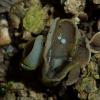
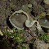
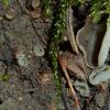
Fruitbody 20-30 mm, cup-like, with violaceous to brown-violaceous hymenium and a pale violett to greyviolett, rough outside, gives a opalescent "latex" after breaking it.
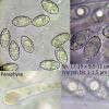
Spores ellipsoid to distinct fusiform, with distinct warts 1-1,5 µm high, even more conspicuously (broader, not higher) at the poles (see photos), with only one vakuole, sometimes with one big and some smaller ones, 17-19 x 9,5 -11 µm.
Paraphyses cylindrical, with swollen apex, septate, in the apex filled with brown-orange material, 3,6 µm, apex up to 5 µm broad.
Found on soil, between moss in a Fagus-forest.
Nicolas VAN VOOREN,
13-10-2010 21:13

Re:Peziza spec.
This is Peziza succosa.
Björn Wergen,
13-10-2010 23:03

Re:Peziza spec.
sure? The "two" species grew together and were definitely distinguishable from each other by different colous and fruitbody-size...I had lots of succosa collections this year to see the difference to this species here, especially the exceptional formed spores (nearly fusiform) and the broader warts at the poles...
cheers, kazuya
cheers, kazuya
Nicolas VAN VOOREN,
15-10-2010 07:57

Re:Peziza spec.
P. succosa offers a great variabilty in its macrsocopic aspects. Your first photograph is typical: white latex, becoming yellowish when drying, colour of hymenium, colour of the external surface. The microscopic features are also in accordande with this species.
Is it possible to see the spores in CRB?
Is it possible to see the spores in CRB?
Björn Wergen,
18-10-2010 14:02

Re:Peziza spec.
Hi Nicolas,
its not possible for me to see spores in CRB, I do not have any chemicals to proof it...
cheers,
Kazuya
its not possible for me to see spores in CRB, I do not have any chemicals to proof it...
cheers,
Kazuya


