
19-05-2026 10:27
Patrice TANCHAUDBonjour, récolte récente sur terre retournée i
04-06-2026 18:39
Gernot FriebesHi,I collected this species in two different locat
22-05-2026 13:29
Gernot FriebesHi,I am curious to hear your opinion on this mater
04-06-2026 10:50
 François Freléchoux
Bonjour, J'ai trouvé hier un petit asco observé
04-06-2026 07:02
François Freléchoux
Bonjour, Voici la description d'une espèce qui p
04-06-2026 13:34
Gernot FriebesHi,I am interested to hear your opinion on this Le
04-06-2026 11:36
Gernot FriebesHi,found on Vaccinium myrtillus.Asci: IKI –, 8-s
18-10-2022 00:12
Valencia Lopez Francisco JavierHola amigos/asRecientemente encontré esta colecci
 Hi all,
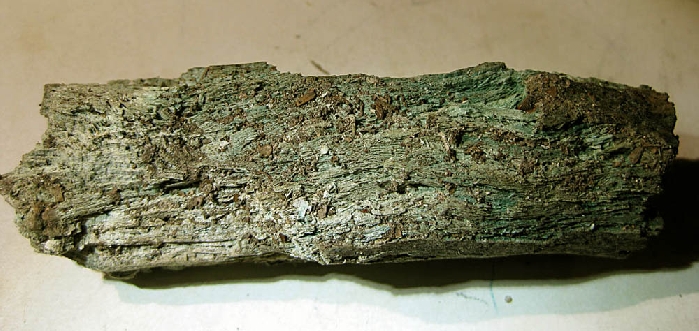
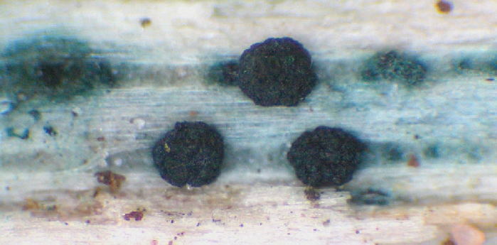
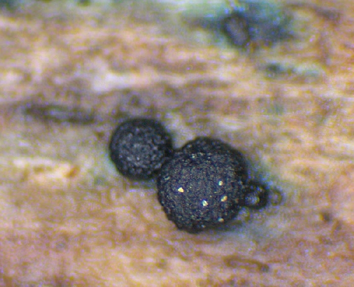
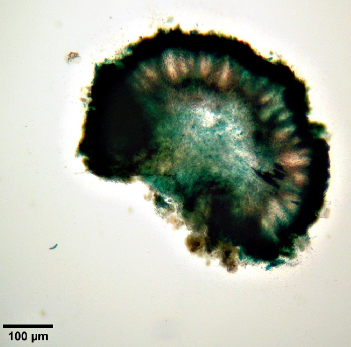
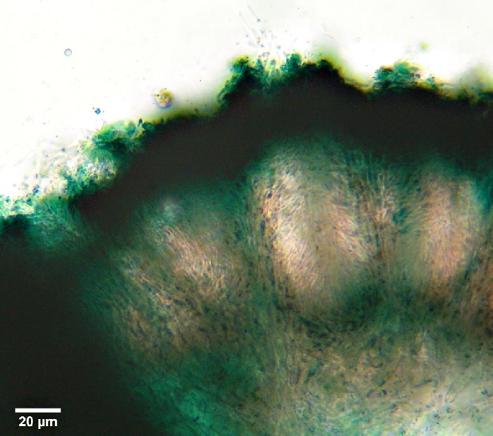
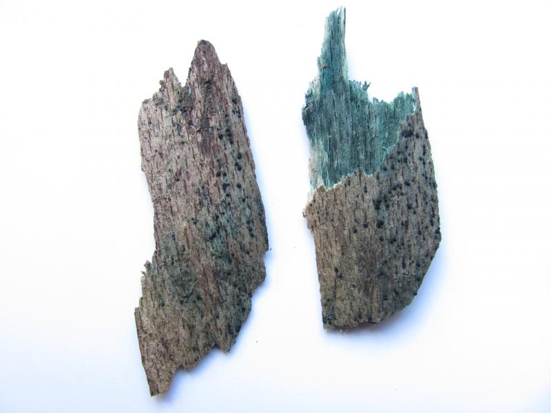
I collected a broken twig of possibly Fagus (according to te remainig bark), stained green by a Chlorociboria. Under the dissecting microscope it featured some "black" fruiting bodies resembling to Bertia moriformis by shape. They appeared always allong the green stained portions of the wood and were soft, gelatinous when cut and featured lots of green pigments as well. Ass. on other parts of this twig were Calocera cornea and a yet unidentified discomycete (my macroscopical guess is a Hyalorbilia).
Can somebody of you give a hint to literature for the anamorph or is this a sclerotium of C.?
Attached are some pictures that could be better.
Best regards,
Martin
my fungus seems to be Dothiorina tulasnei, an anamorphic fungus in discussion to be related to Chlorociboria. Though Sánchez & Bianchinotti (Mycotaxon 102, 395-402) are raising doubts (p.401). Reportedly there was no successful culture made of Ch. yet.
Best regards,
Martin
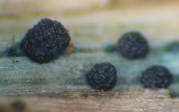
first a little better macro:
Has somebody of you experienced this fungus?
Best regards,
Martin

that's wonderful, and it resembles a bit the Coryne anamorph of Ascocoryne. Never seen. I have this ana-teleo relation in my database, but saw it only in Dixon 1975.
Zotto

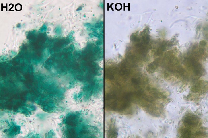
are cells inside of the locule become green in KOH and conidia formed in chains?
If so, then I think your fungus is probably Dothiorina tulasnei.
Regards,
Christian
I did not note a reaction of the green pigment of Chlorociboria in KOH, only a dissolution of the exudate which turned yellowish-greenish, but not staining the medium (Chl. aeruginoa).
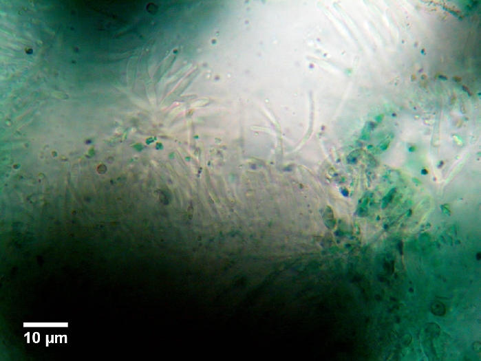
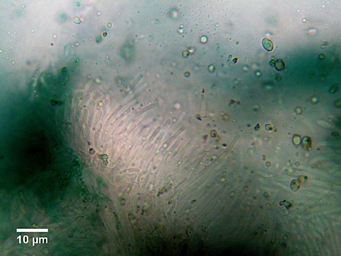
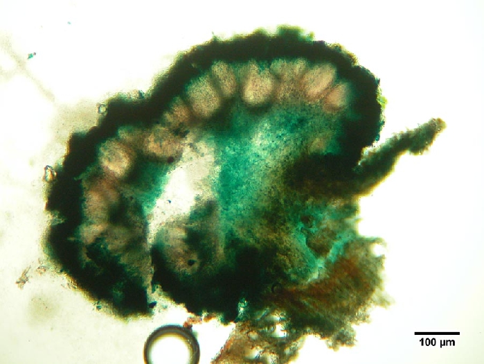
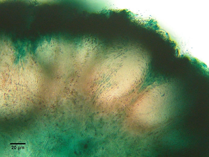
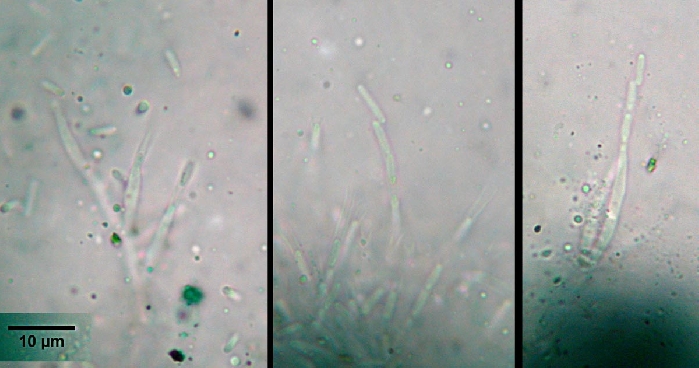
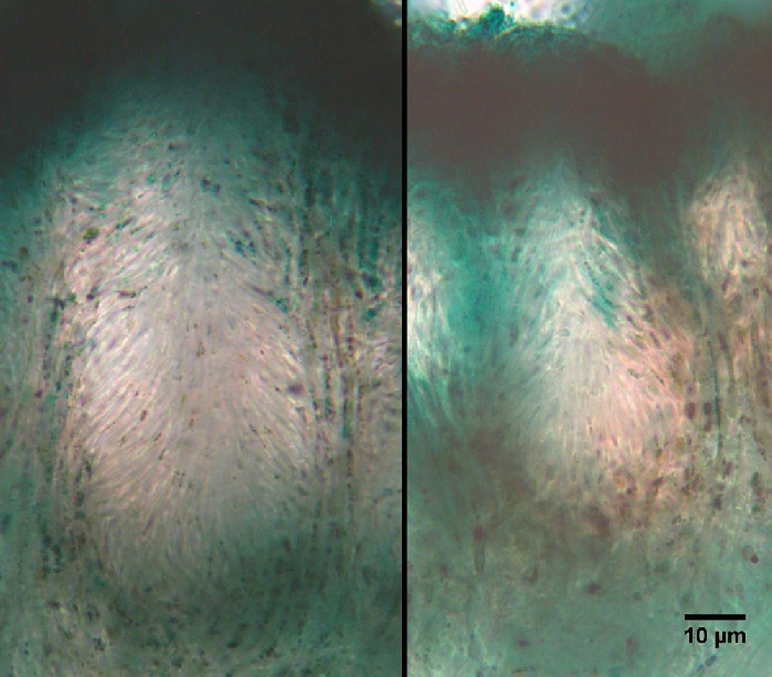
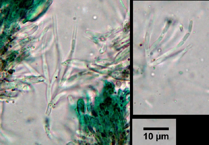
I made a new section in order to see better the inner structure of the loculi. The conidiophores are arranged all along the inner walls with their necks upwards. And yes, Zotto, the conidia are produced in chains inside a collarette. Hope one can see that.
KOH has a bleaching effect turning the bright turcoise into olive-brown.
Best regards,
Martin
I also believe this is Dothiorina tulasnei
I´m sending a pdf file.
best regards
Virginia
 13062.pdf
As I understood from your paper, you don't belive, that Dothiorina is related to Chlorociboria?
Best regards
Martin
best regards
Virginia
Regards
Martin
Does anybody have a pdf of this paper?
Johnston PR, Park D. 2005. Chlorociboria (Fungi, Helotiales) in New Zealand. New Z. J. Bot. 43:
679-719.
Zotto
You can download here:
http://www.royalsociety.org.nz/publications/journals/nzjb/2005/043/
Grüße,
Martin
EDIT: this may work...
The substrate wasn´t stained bluegreen, but logs with Chlorociboria were stained bluegreen (we have also seen Chlorociboria on the same host).
I´m sending you the paper to your mail
Virginia

Dear Friends
Among the samples from the Ukrainian Carpathians I also found Dothiorina tulasnei. The fungus formed sporulation on fallen decorticated branches of Fagus, which were coloured by Chlorociboria (see photo below).
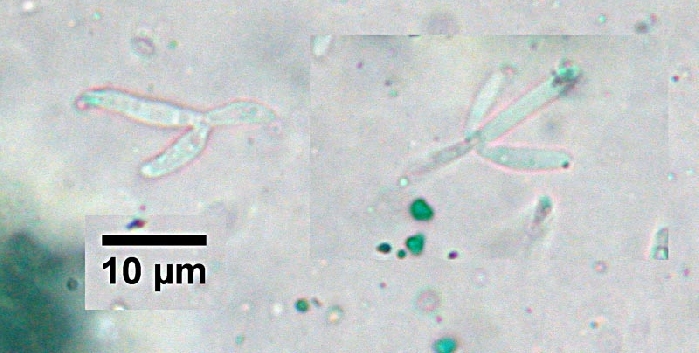
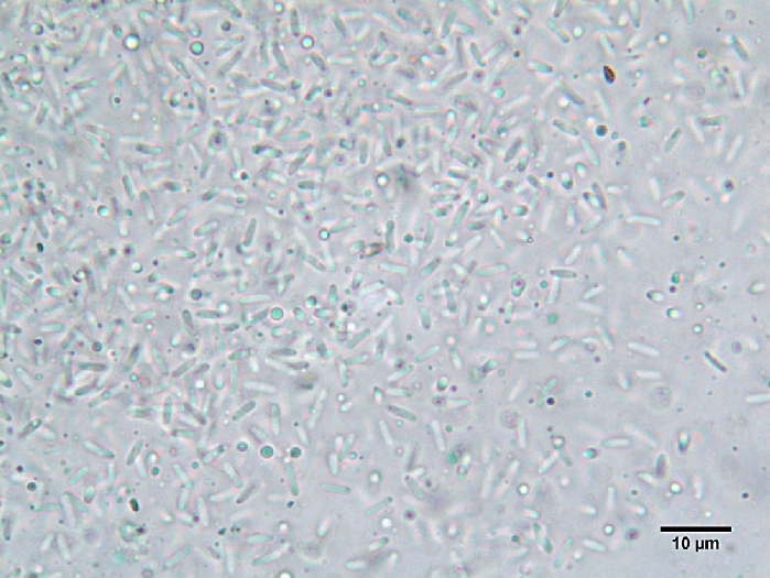
In my sample, the conidia are slightly different in size from those that have been described by Virginia. Their length is 3,4-5,6 (6,5), width - 0.9-1.1 (1.2) micron.
If anyone interested, I could send samples for more in-depth research.
Thank you for the wonderful discussion on the forum!
It helped me to understand with what I am dealing with.
Alex
wonderful collection! Keep it aside! I am still waiting for a branch with both the ana-and teleomorph together as in the Tulasnes plate... ;-)
Cheers,
Martin