13-02-2026 03:30
Hello! I found these immersed perithecia on a stic

13-02-2026 18:05
Margot en Geert VullingsOn February 9, 2026, we found these small hairy di

12-02-2026 21:34
patrice CallardBonjour, la face infÃĐrieure des feuilles ce certa

11-02-2026 22:15
 William Slosse
Today, February 11, 2026, we found the following R

12-02-2026 14:55
Thomas LÃĶssÃļehttps://svampe.databasen.org/observations/10581810

11-02-2026 19:28
 Lothar Krieglsteiner
on small deciduous twig on the ground in forest wi

25-04-2025 17:24
Stefan BlaserHi everybody, This collection was collected by JÃ

10-02-2026 17:42
 Bernard CLESSE
Bonjour à toutes et tous,Pourriez-vous me donner
 Has Sphaerosporella brunnea in the hymenium the same hairs than margin ones?
I am sure it is not a contamination. The hymenial hair are not abundant but constant.
Miguel Ã. Ribes

I haven't treated any Sphaerosporella for a long time (though my associate and me are trying to come accross one for a years to make detailed study) but when I just browsed my old few collections of S. brunnea form 90-ties I saw no data on such hairs (only normal ones!). Also these hymenia were not so rich in carotenes as yours. It was sandy- to yellowish-grayish ochre. Sometimes also with olivaceous tinge. Do you have ascospore diameter and hair (length/width) measurements?
Cheers,
Neven Matocec
Je n'ai jamais rencontrÃĐ de S. brunnea avec une couleur orange, mais... !
Dommage, comme souvent dans les demandes formulÃĐes sur le site, il manque une description du champignon qui permettrait de renseigner de maniÃĻre plus de pertinente. Par ailleurs, je ne suis pas certains qu'il s'agisse rÃĐellement de poils, mais de paraphyses ÂŦ anormales Âŧ ceci d'aprÃĻs le contenu protoplasmique qui semble identique aux paraphyses normales. Il conviendrait ÃĐgalement de savoir si ces ÃĐlÃĐments, de type acuminÃĐ, apparaissent ÃĐgalement parmi l'hymÃĐnium des jeunes sporophores ou si, au contraire, ils sont prÃĐsents parmi l'hymÃĐnium de sporophores ÃĒgÃĐs !?
Cordialement
RenÃĐ
?

I recently found an ascomycetes (without fire, hidden under a pile of branches) which I have determined as Sphaerosporella brunnea. This was very similar to the genus Trichophaea (gray-white fruit layer, max. 5 mm) but with round spores, and the sparse hair. Microscopically, there was apparently no difference to Sphaerosporella brunnea. Now when I see the image of Miguel, I think there must be two species or Sphaerosporella brunnea is highly variable.
Since we have to stay tuned to explore that.
Peter

Sorry at all. I did not made yesterday a complete description because I had no time. Sorry again.
Here it is:
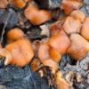
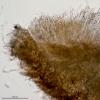
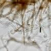
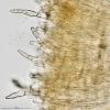
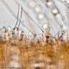
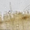
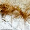
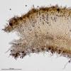
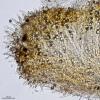
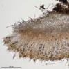
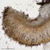
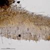
Macro: In fired wood. Apos 3-5 mm broad, sesil, convex orange hymenium, margin and lateral sides with dark and short hairs.
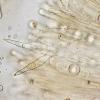
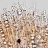
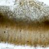
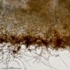
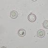
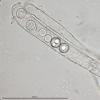
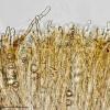
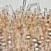
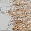
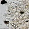
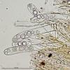
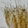
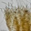
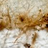
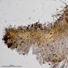
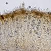
Micro: Cylindrical asci, with croziers, no reaction with IKI. Sphaerical spores. Ectal excipulum angular, becoming cylindrical at the margin. Margin with pointed hairs, hialine, 2-3 septa. Sides with rounded or pointed hairs, light brown, longer than marginal hairs. Base with cylindrical subiculum. Hymenium with hairs Âŋ? like margin ones. Paraphysis slightly thickened at apex.
And this are the measurements:
Spores (1000x, water, fresh)
(12.5) 14 - 16.1 (16.8) x (12.1) 13.7 - 15.9 (16.3) Âĩm
Q = 1 - 1.05 (1.1) ; N = 54
Me = 15.2 x 14.9 Âĩm ; Qe = 1
Asci (1000x, water,fresh)
(212.4) 218.6 - 250.3 (256.4) x (18.5) 18.8 - 22.8 (24.8) Âĩm
N = 10; Me = 230.5 x 21.1 Âĩm
Hymenial hairs (600x, water, fresh)
(64.7) 68.4 - 87.2 (103.2) x (6.4) 7.2 - 9.9 (11.3) Âĩm
N = 22; Me = 76.9 x 8.5 Âĩm
Margin hairs (600x, water, fresh)
(50) 61.8 - 92.8 (96.9) x (5) 6.2 - 10.7 (12.9) Âĩm
N = 41; Me = 73.3 x 8.4 Âĩm
Lateral hairs (600x, water, fresh)
(73.6) 107.9 - 171.3 (185.8) x (3.9) 5.4 - 8.8 (10.1) Âĩm
N = 22; Me = 138.3 x 7.1 Âĩm
Width paraphysis apex (600x, water, fresh)
(5.4) 6.2 - 7.4 (8.1) Âĩm
Me = 6.7 Âĩm
Thank you.Â
Miguel Ã. RibesÂ
Merci pour les complÃĐments apportÃĐs à la description de ce champignon.
Nous sommes à prÃĐsent certain que nous avons une espÃĻce appartenant au genre Sphaerosporella.
J'ai consultÃĐ les publications de divers auteurs, dont Seaver et Rifai. Aucun des Auteurs ne fait ÃĐtat de poils ou de paraphyses simulant des poils dans l'hymÃĐnium, qui s'agisse de S. brunnea et de S. hinnulea, cette derniÃĻre ÃĐtant dÃĐsignÃĐe comme distinct de S. brunnea, ce qui, Ã mon avis n'est vraiment pas certain.
J'ai eu l'occasion d'ÃĐtudier S. brunnea rÃĐcoltÃĐ une place à feu (saprophyte), ainsi que sur pourtour (formÃĐ de terre enveloppÃĐ de textile) de plantules de Ilex (mycorhize). Je n'ai pas observÃĐ cette particularitÃĐ. Je vais ressortir les exsiccata et contrÃīler à nouveau, mais ceci durant l'hiver. Si je devais trouver ce genre de cellules dans l'hymÃĐnium, j'en ferai ÃĐtat sur le site.
Pour l'heure, je pense qu'il s'agit d'une anomalie. A ce propos, vous n'avez pas rÃĐpondu à la question qui ÃĐtait de savoir si les paraphyses ÂŦ anormales Âŧ se trouvaient prÃĐsentes à la fois sur les exemplaires jeunes et sur les exemplaires ÃĒgÃĐs ? Il arrive en effet que certaines paraphyses continuent de croÃŪtre et se dÃĐforment avec l'ÃĒge.
Cordialement
RenÃĐ
?

I have study 3 small apos (2 mm diam.) and the hymenial hairs are more and more abundant than in big apos. Hymenial hairs are more abundant near the margin than in the center. Perhaps young apos are full of hairs, in the margin as in the hymenium; and when they developed, are more and more differenciated and only keep hairs in the margin and in the nearest margin hymenium?
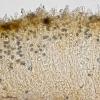
New pictures of young apos.
Very kind RenÃĐ, Neven and Peter,
Miguel Ã. Ribes

ÂĄCurioso lo de los pelos en el himenio! Esto demuestra que no hay mucha diferencia entre las cÃĐlulas del margen y las parÃĄfisis. Simplemente que se han "desorientado" y han preferido crecer y diferenciarse en el centro del apotecio en vez de en el margen.Â
ÂŋHas mirado si las esporas maduras presentan ornamentaciÃģn?, tal y como nos muestra Guy G. en este mensaje:Â http://www.ascofrance.fr/search_forum/13328
Strange those hairs in the hymeniun! This proves that there is not much difference between marginal cells and paraphyses. Just these ones have "lost" their normal position and have prefered to grow and differentiate in the hymenium instead at the margin. Have you seen if mature spores develop ornamentation as Guy G. shows us in this message: http://www.ascofrance.fr/search_forum/13328
Un saludo a todos,
RaÚl

SÃ, esos pelos estÃĄn completamente desorientados, pero son muchÃsimos y en todos los ejemplares.
Acabo de hacer nuevas preparaciones para ver la posible ornamentaciÃģn de las esporas y son completamente lisas, ni rastro de ornamentaciÃģn, y ningÚn parecido a la del enlace que envÃas de Guy.
Me resulta extraÃąo que nadie haya visto antes estos pelos en esta especie, porque no pasan desapercibidos, son muy abundantes.
__________
Yes, those hairs are completely disoriented, but there are many, and in all apos.
I just made new preparations to see the possible spores ornamentation, and they are completely smooth, no trace of ornamentation, and no resemblance to the Guy's link you send.
It is strange nobody has seen these hairs in this species, because the are obvious and very abundant.
Saludos,
Miguel Ã. Ribes

Thanks for checking the ornamentation. Never found this species. If I do, I will send you the micro.
Saludos,
RaÚl
Je me permets de revenir sur la particularitÃĐ observÃĐe sur cette rÃĐcolte de S. brunnea, la terminaison ÂŦ piliforme Âŧ de certaines paraphyses.
Comme je l'avais indiquÃĐ dans mon prÃĐcÃĐdant message, du 16 octobre, il arrive que certaines paraphyses continuent de croÃŪtre en se dÃĐformant avec l'ÃĒge.
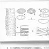
Le hasard faisant parfois bien les choses ! J'ai, en recherchant un article dans l'ouvrage de RIFAI (1967) The Autralasian Pezizales in the Herbarium of the Royal Botanic Gargens Kew, trouvÃĐ un dessin reprÃĐsentant des paraphyses de Cookeina sulcipes, dont l'une porte une cellule clavÃĐe, septÃĐe, à paroi ÃĐpaisse, qui contraste fortement avec le sommet des autres paraphyses (voir annexe). Dans la description, Rifai indique que de telles clavules sur les paraphyses sont frÃĐquentes. J'ai retrouvÃĐ dans la description de LE GAL (1953) Les DiscomycÃĻtes de Madagascar, ces mÊmes ÃĐlÃĐments au sommet de certaines paraphyses. Il semble donc, pour cette espÃĻce, que cette ÂŦ anomalie Âŧ soit devenu une particularitÃĐ spÃĐcifique.
Pour ce qui est de S. brunnea, je suis convaincu qu'il s'agit d'une anomalie du mÊme type que l'on peut rencontrer chez C. sulcipes et chez d'autres discales. Il s'agit d'une de ces anomalies qui font que, lentement, les espÃĻces se modifient et ÃĐvoluent.
Cordialement
RenÃĐ
?

Thank you again. The results of your research are very interesting. I have not Rifai's work, but I have Le Gal's one, and actually describe this feature of some paraphysis in Cookeina sulcipes and Pseudoplectania nigrella.
An interesting finding. Perhaps S. brunnea is evolving in Cantabria into "S. himenopilosa"!!!
Best regards,
Miguel Ã. Ribes
