
26-05-2026 22:44
Ethan CrensonHi all, I think I have Incrucipulum capitatum her
26-05-2026 21:25
Dirk GerstnerHello everyone, I'm completely stumped by this li
22-05-2026 14:44
 Lothar Krieglsteiner
in unripe condition citrine yellow, then soon fadi
25-05-2026 16:44
François BartholomeeusenHi forum members,During an excursion organised by
23-05-2026 11:44
 Charles Grapinet
Hello, I am having trouble identifying this copro
25-05-2026 16:35
 Bernard CLESSE
Bonjour à toutes et tous,J'ai trouvé récemment,
22-05-2026 13:29
Gernot FriebesHi,I am curious to hear your opinion on this mater
23-05-2026 18:57
Sylvie Le GoffBonjour à tousRécolté sur une branchette de Sal
22-05-2026 21:35
Steve ClementsBonjour, I expected this find on old wood on our

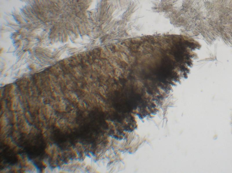
Hello, dear friends!
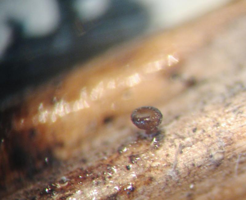
These 2 specimens some time ago i identidied as C. cyathoidea. Now I see some differences in spore morphology, and I wonder whether one of them could be C. pallida.
The 1st was examined in fresh condition, the 2d in exciccated state.
So, the 1st specimen was collected in oak forest, on Urtica dioica rotten stem.
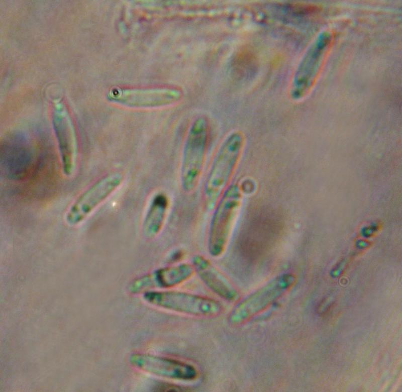
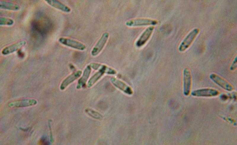
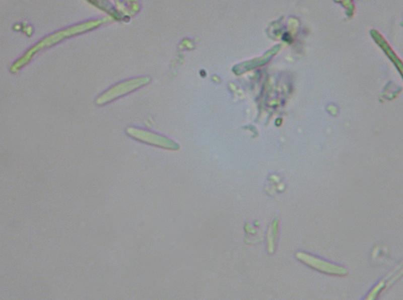
Spores 7,3-12,6*2,2-3,6 um, with 1-3 small oil drops on each end.
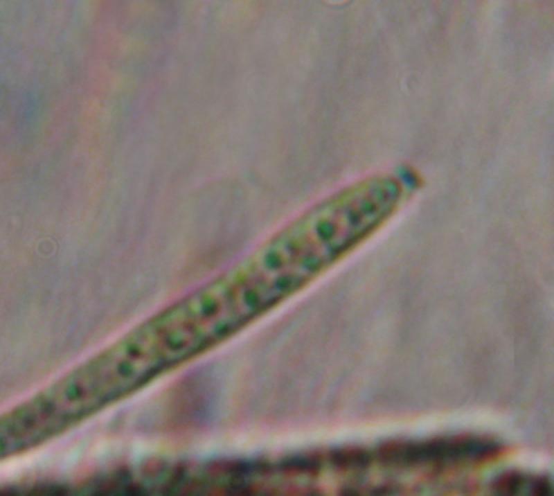
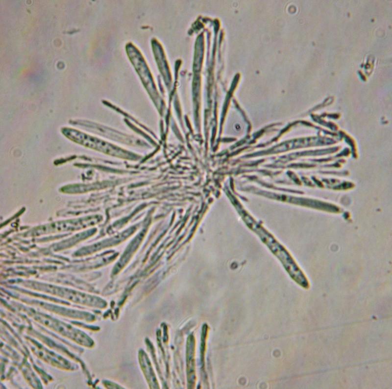
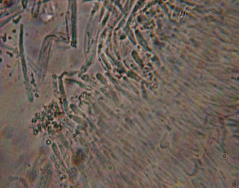
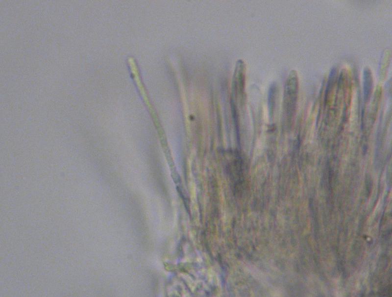
Asci IKI B, with croziers, 49-68*3,6-5,5 um
Cheers,
Irina

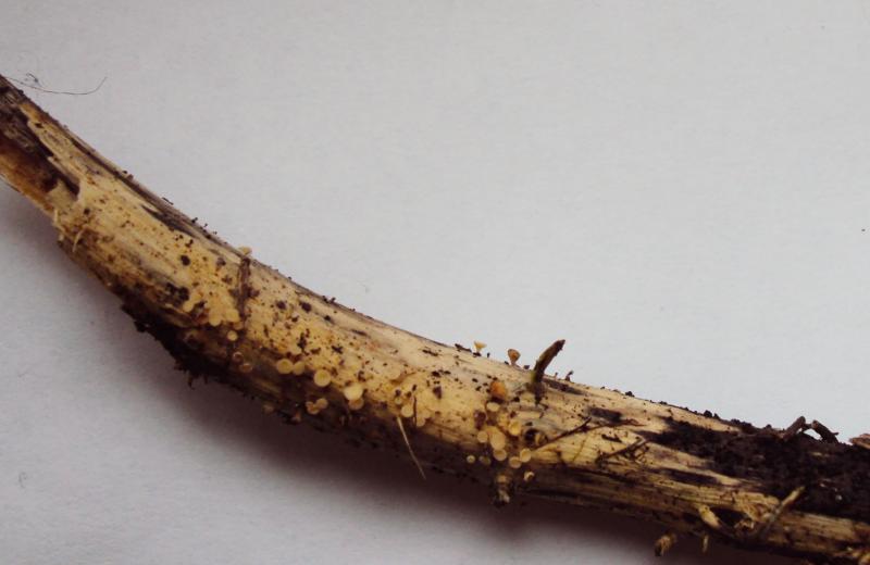
The 2d one was collected on a meadow, on rotten herbaceous stem.
Spores 9,1-12,7*1,8-2,7 um, fusiform, sometimes slightly S-shaped
Asci IKI B, with croziers, 44-60*3,8-5,5 um


in order to confuse you a bit :-)
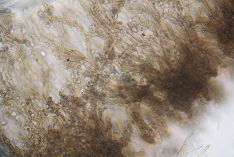
C. pallida is a species with marginal teeth, at least as I understand it. It was treated by Breitenbach & Kränzlin under the wrong name C. dolosella. The marginal teeth are not shown on their photo, but they are mentioned, and I reexamined their material:
your whitish specimen could well be C. cyathoidea, quite a variable species. Are the spores actually up to 3.6µm? Regrettably, only the spores are alive in your preparations. Maybe you press too strong. The apical ring photo seems to exclude hymenoscyphus repandus.
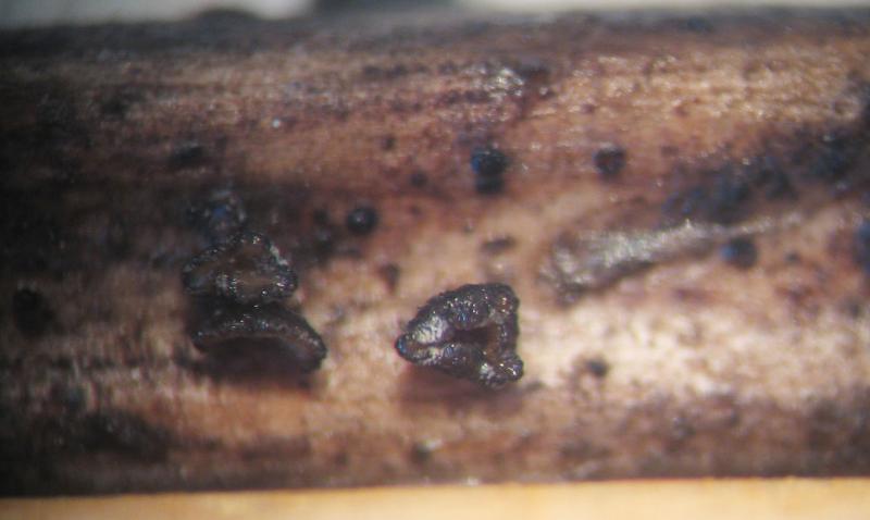
The brown one reminds me of C. cacaliae.
Zotto

Hello, Zotto!
And thank you for answer.
Yes, I know about marginal teeth in C. pallida, but in my opinion they probably could be poorly visible/destructed, so on. The 2d one was collected in dry condition, so I cannot say surely whether it was brown in living state or not.
With best regards,
Irina