
28-05-2026 16:15
James MitchellHello,Does anyone have the original publication of
28-05-2026 11:06
Thomas Læssøehttps://svampe.databasen.org/observations/10596750
23-05-2026 11:44
 Charles Grapinet
Hello, I am having trouble identifying this copro
25-05-2026 16:44
François BartholomeeusenHi forum members,During an excursion organised by
26-05-2026 21:25
Dirk GerstnerHello everyone, I'm completely stumped by this li
26-05-2026 22:44
Ethan CrensonHi all, I think I have Incrucipulum capitatum her
22-05-2026 14:44
 Lothar Krieglsteiner
in unripe condition citrine yellow, then soon fadi
25-05-2026 16:35
 Bernard CLESSE
Bonjour à toutes et tous,J'ai trouvé récemment,
22-05-2026 13:29
Gernot FriebesHi,I am curious to hear your opinion on this mater
23-05-2026 18:57
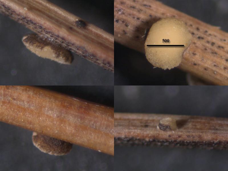
Sylvie Le GoffBonjour à tousRécolté sur une branchette de Sal
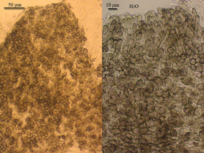
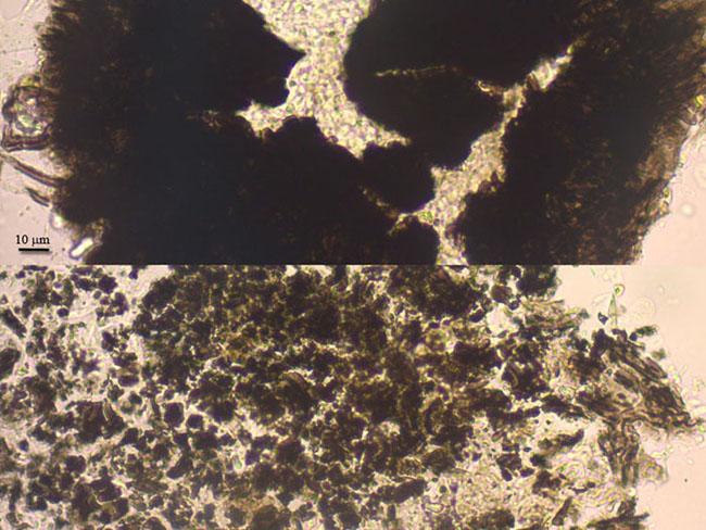
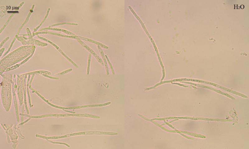
It is growing on litter of Eriophorum vaginatum in bog.
Apothecia cupulate, sessile, arise from under scutulum, young hemispherical with small opening, later cupulate with incurved margine, to 650 mk wide, 150 mk high; hymenium surface yellowish, convex; outer surface brown to pale brown, rough from loose cells of outer layer, edge lighter; shield dark brown, ellipsoid, concave, about 240 mk in diam; base with some hyphae attaching to the substrate.
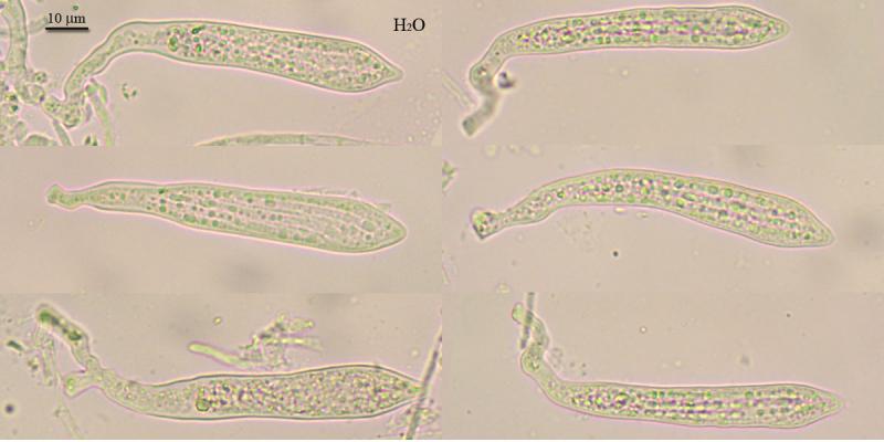
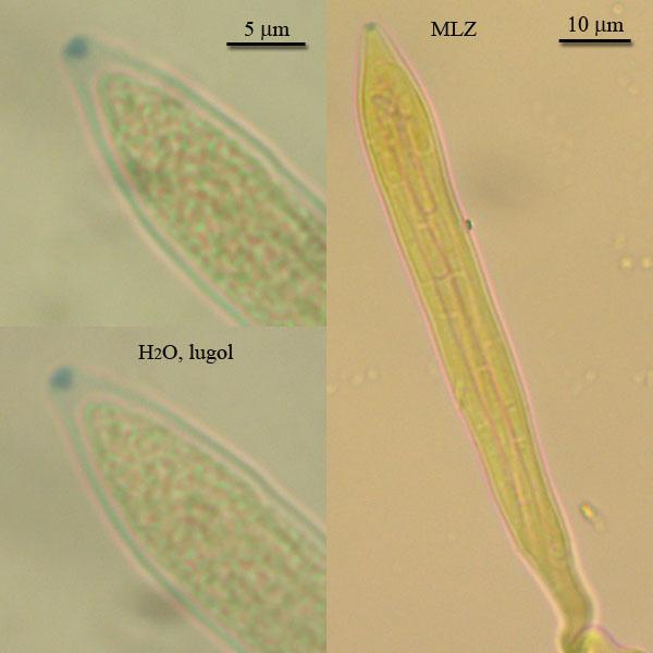
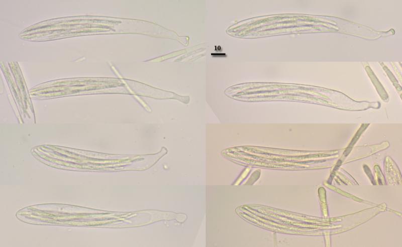
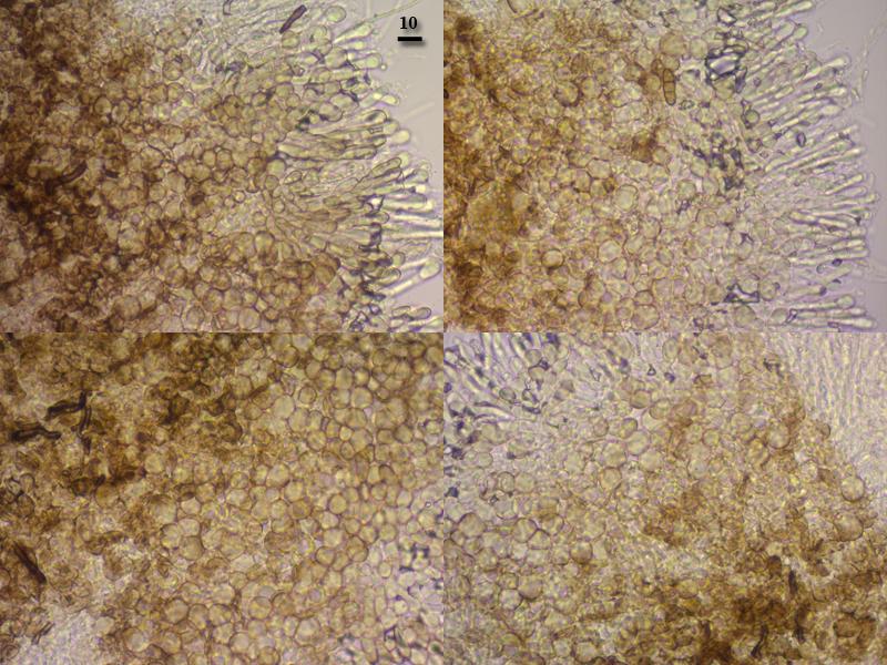
Excipulum from medullary and ectal layers, medullary from not gelatinose, loose, intervowen hyphae about 1,7 mk thick, their tips form ectal- layer, e.g. end cells (2-3) of hyphae enlarged, densely placed, outer cells covered by brown exudate and measured about 12,5 x 9,5 mk, at the edge of receptacle the same cells have hair-like shape, e.g. cylindrical, with clavate ends (hair length about 50 mk, width at upper cell 5 mk); shield fomed by glued dark brown hyphae of indistinct arrangement; asci enlarged to conical tip, long or short stalked, occationally two-stalked, with clamp, wall thickened in upper part, pore euamyloid, trapeziform, 75-95 x 8,4-13; paraphyses cylindrical, slightly enlarged to tip (2-3 mk), branched, segmented, with oily content in single segments (revived in water); spores vermiform, broader and 2-3 segmented in upper part, narrower in basal part, obtuse- ended (or some equally broad, obtuse and attenuated at ends), with many round and ellipsoid guttules (rehydrated in water), 53 (39-66) x 2,8 (2,2-3,5) (N=15).
N61,063892° E69,455695°, 06.08.2012.


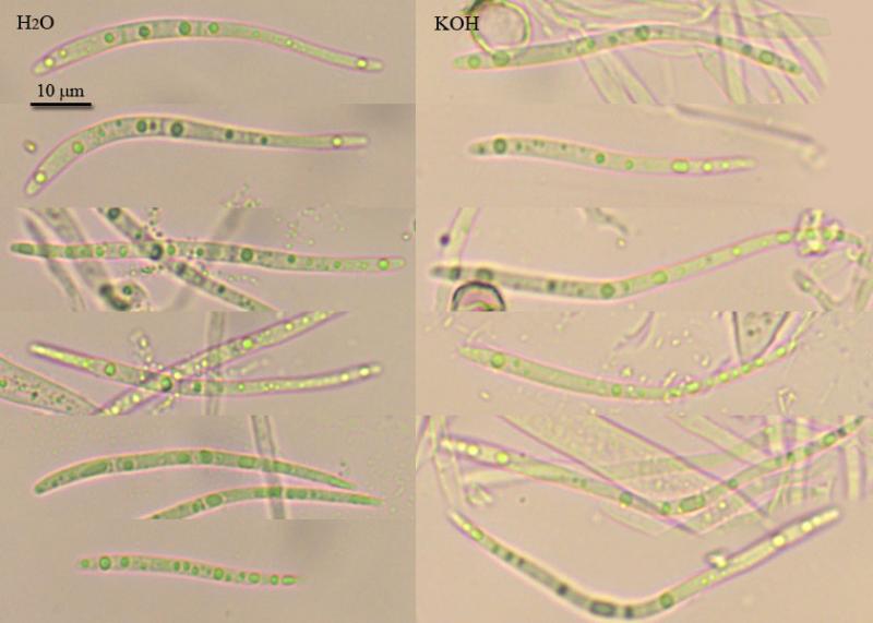

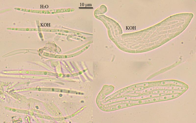
Perhaps also the ascus shape (kind of conical apex) suggests that your specimen is not B. iridis. Who knows... The Scutomollisia species that I know do have these VBs, and partly these give a distinct KOH-reaction. You did not test what KOH evokes at the moment when it comes in contact with the paraphyses?
Zotto

Stip


I have restudied the Crouan type (HB 6604). Under the below link you find all my images on iridis:
https://www.cubby.com/p/_778a6f3587954e008790666f822f8e4e/7a+Helotiales#7a%20Helotiales/Mollisiaceae/Dennisiodiscus
Yes, iridis tends to be thicker in the middle, but sometimes it is cylindrical and tapers only close to the ends or only at the base.
Le Gal's drawing shows also narrowly fusiform spores up to 60 µm length.
Zotto
LeGal's description seems to be a bit strange with asci 180-220 long, although it fits her drawings, so this is a different species from the one presented here. In contrast C. Scheuer's Scutomollisia spec indet. (Bibl Mycol 123 p. 169) seems to be a perfect fit.
Stip
There are several things strange in Le Gal's redescription: apos she found 2-3 mm while I only up to 1 mm. My asci were only 120-140 x 8-9 while she wrote 180-220 x 8-10. I saw no free spores but many inside asci. It is possible that apothecia studied by her were more mature and actually show such differences.
Zotto
unfortunatelly, i had not paid attention to vital paraphyses, there are pictures taken in vital below, but probably i added KOH to it too (...) So, it is impossible to say about VBs now, only if it will collected again (it was collected twice, so there is chance).
Inspite of large opened apos they seem underdeveloped, only in few of them asci contained spores, and all were inside. Those spores with one enlarged end i suppose to be developed more fully. But majority of spores difficult to extract from asci, they tend to break apart, and have ends similar in width. Some spores have empty ends for some reason. There one more picture of spores in water and KOH, and tapped asci with spores.
What about underdescribed taxons and riddles, i hope that there are will be possibility for some collaborative work with you in future. Now i will proceede with working out other specimens. I think for me at this stage more usefull to know different groups and to identify in prior all previous year collection. And to start looking more attentively in next season (I am supposed to finish my phd work in next year).
Stip, thanks for the paper, it wil usefull many times since there are more collections from bog graminoids.
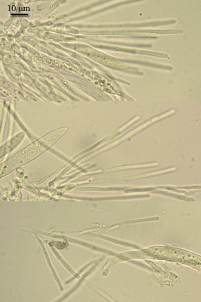
Apothecia arise from scutulum (which about 200 mk broad), cupulate, sessile, hymenium when mature becoming concave, yellowish, outer surface brownish, 600–1000 mk broad, near 200 mk high.?
Excipulum from round cells about 10 mk broad, edge from several rows of prismatic and end clavate cells, brown; asci clavate, with crozier, 98–123 x 13–16.5 (n=10); paraphyses some enlarged to obtuse tip, not or rare branched, filled with many small round (easily collapsing into several elongated) vacuoles, 108 x 5 (upper part) x 2.6 (base) (n=10); spores vermiform, slightly heteropolar, overmature septate, with many tiny oils, 73.3 (65.2–88.2) x 2.8 (2.5–3) (n=15).


Zotto