
09-06-2026 18:32
Camille MertensSur morceau de roseau immergûˋ 0,5 - 0,7 mm de dia
08-06-2026 10:16
 Spooren Marco
I don`t have a clou about this fungus,it is not in
08-06-2026 17:00
FranûÏois BartholomeeusenGood day everyone,ô On June 5 2026, I collected de
07-06-2026 15:10
 William Slosse
Hello everyone,On 05-06-26, I found following asco
05-06-2026 11:02
Thomas LûÎssû¡ehttps://svampe.databasen.org/observations/10596691
07-06-2026 12:09
 FranûÏois Frelûˋchoux
Bonjour, Voici une brû´ve description de ce qui m
07-06-2026 12:43
Steve ClementsBojour. This was a strange find on a stick on my
12-07-2015 00:05
 Nedim Jukic
This one from the same locality as the previous on
06-06-2026 17:44
Steve ClementsBonjour, This disco was on planed wood 3 x 1.5 cm
Hymenobolus agaves anamorph
Miguel ûngel Ribes,
12-03-2013 00:39
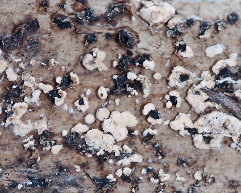
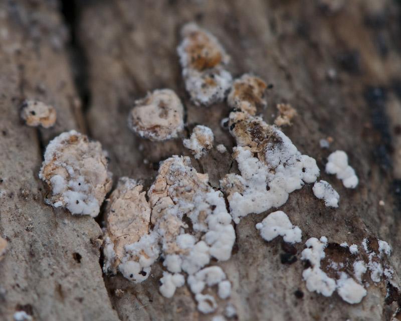
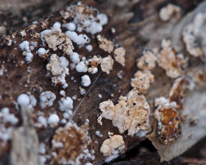
 Good night
Perhaps someone remember this Hymenobolus agaves: http://www.ascofrance.fr/search_forum/10909
Rubûˋn has foung more collections in another Canary Island, La Gomera. In some collections, between H. agaves apothecium, are growing too a white-orange anamorph, 2-5 mm broad, relatively hard (it is posible to cut it).
Is it posible the anamorph of H. agaves? How to study this anamorph?
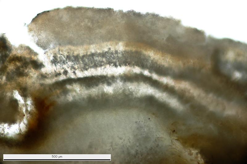
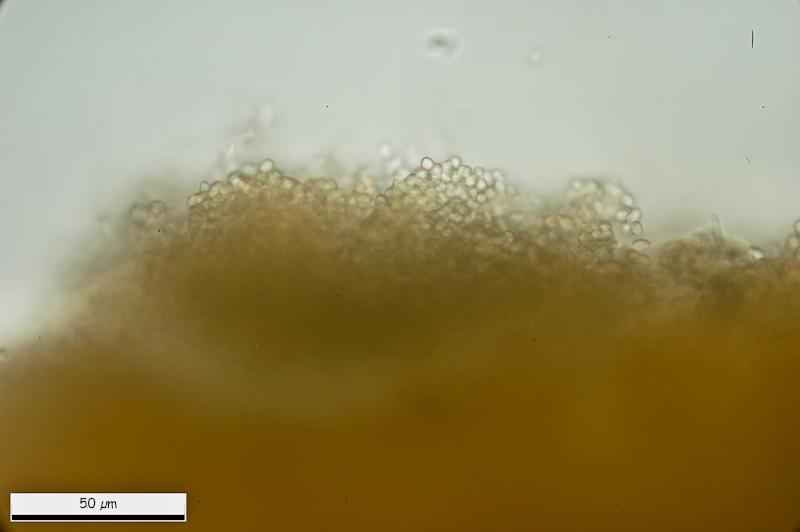
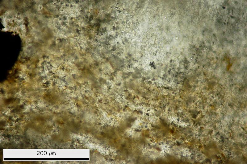
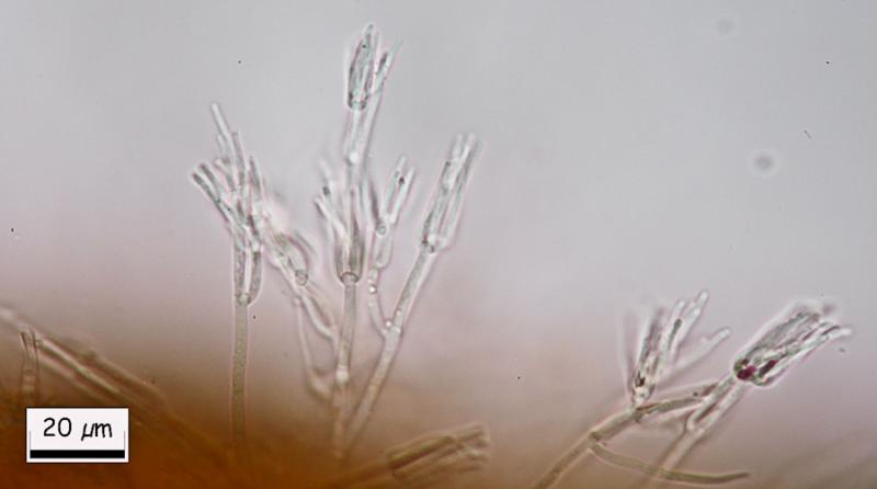
This are general views.
Thank you.
Miguel ûngel Ribes,
12-03-2013 00:44
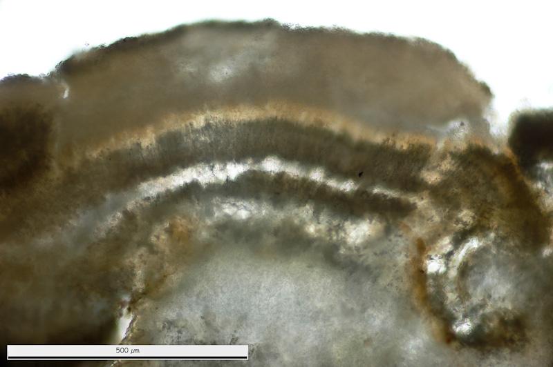
Re : Hymenobolus agaves anamorph
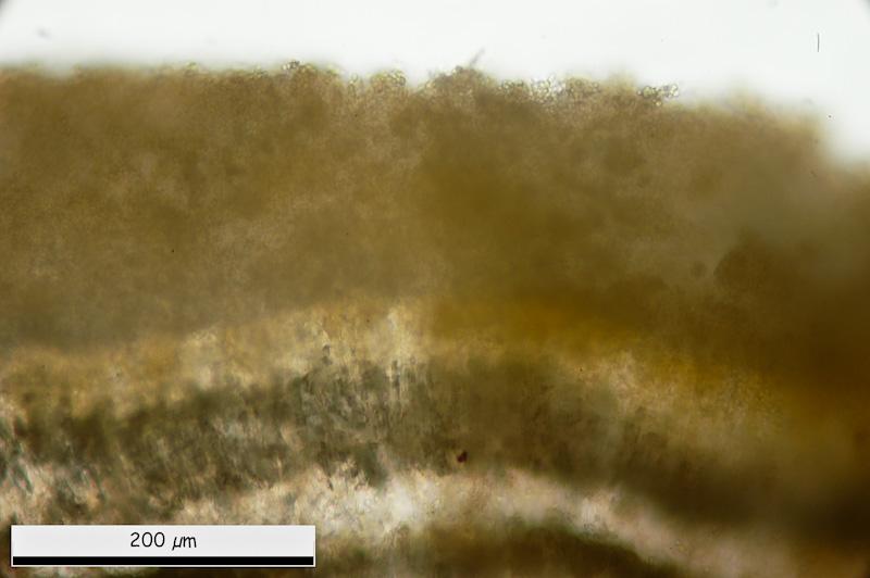
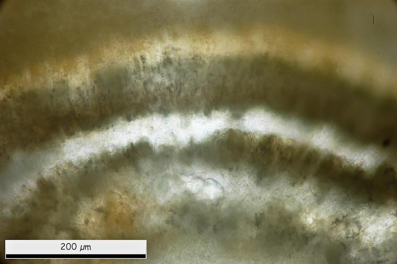
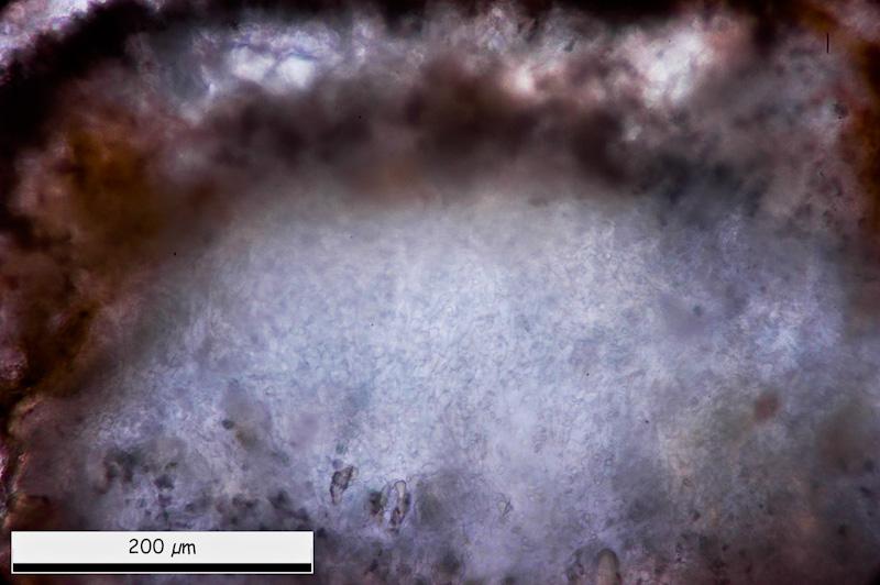
General micro views. It is posible to see some layers. External one with more-less rounded cells.
Miguel ûngel Ribes,
12-03-2013 00:50
Re : Hymenobolus agaves anamorph
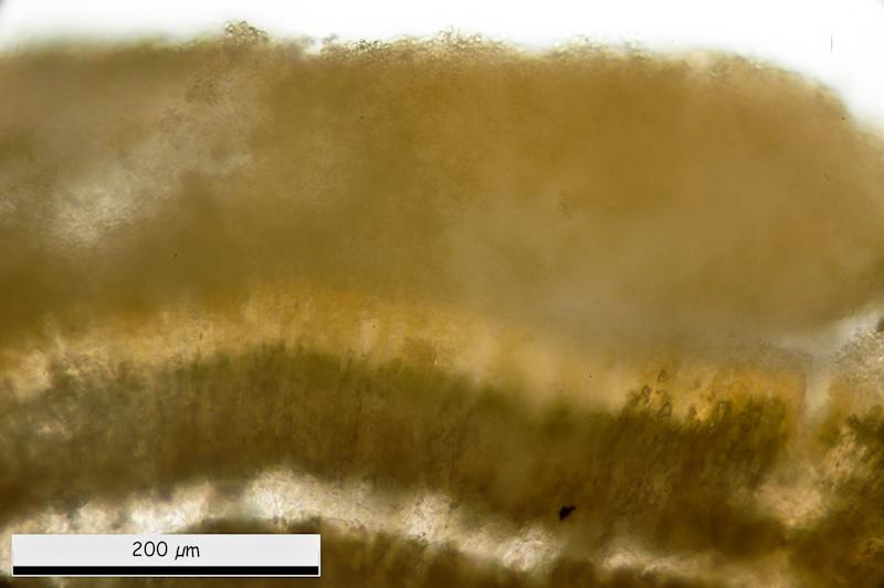
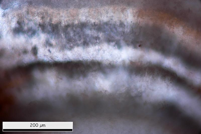
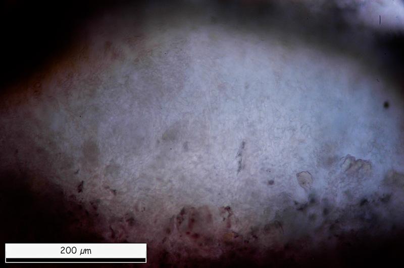
Inside, tow black lines. And in medular area a white area with globose-angular structure mixed with cilyndrical cells.
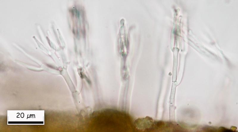
Miguel ûngel Ribes,
12-03-2013 00:53
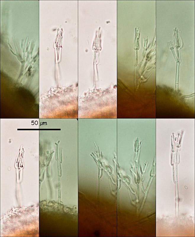
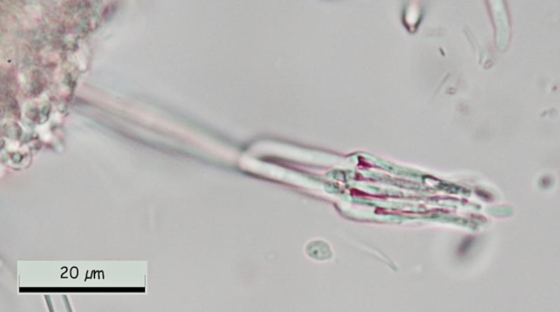
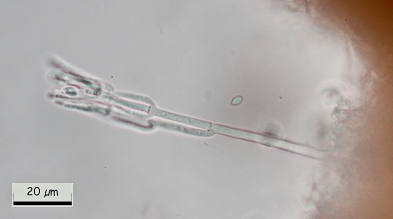
Re : Hymenobolus agaves anamorph
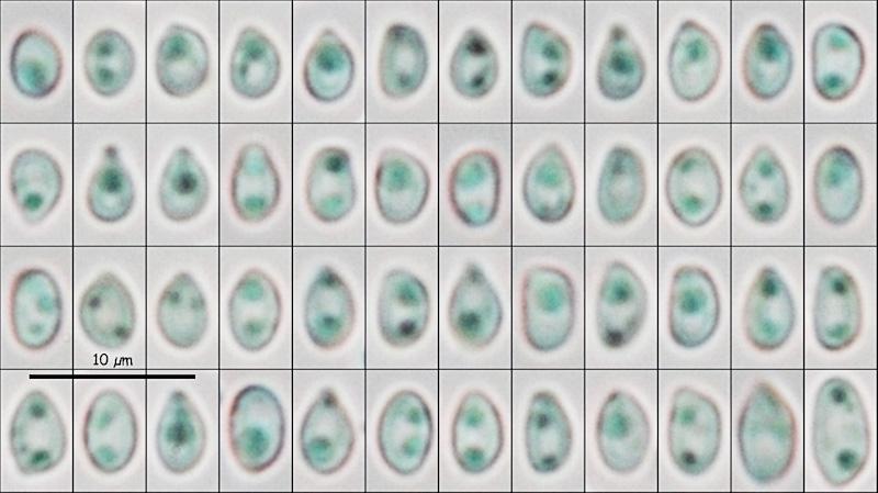
Eliptical conidiospores in water:
(4.06) 4.49 - 5.21 (6.63) x (2.87) 2.94 - 3.35 (3.57) ôçm
Q = (1.28) 1.40 - 1.70 (1.86) ; N = 52
Me = 4.88 x 3.14 ôçm ; Qe = 1.56
(4.06) 4.49 - 5.21 (6.63) x (2.87) 2.94 - 3.35 (3.57) ôçm
Q = (1.28) 1.40 - 1.70 (1.86) ; N = 52
Me = 4.88 x 3.14 ôçm ; Qe = 1.56
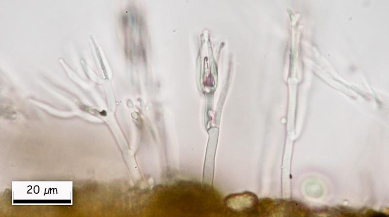
Miguel ûngel Ribes,
12-03-2013 00:55
Re : Hymenobolus agaves anamorph
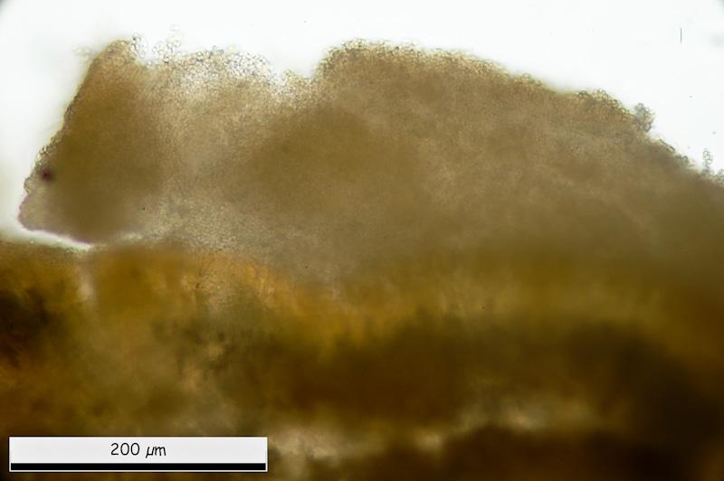
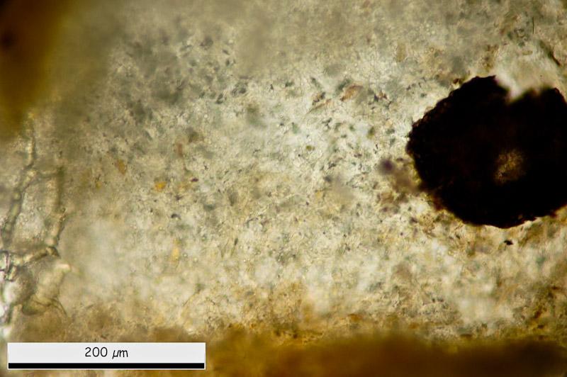
And finally, at the margin, this conidial structure.
Than you in advance.
Miguel û. Ribes
Than you in advance.
Miguel û. Ribes
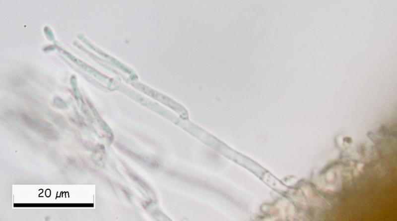
Hans-Otto Baral,
12-03-2013 08:06

Re : Hymenobolus agaves anamorph
Great, Miguel! Could you please show us a closeup of the conidiogenous cells, were the conidia emerge? I assume they are phialidic. Then we can search in Genera of Hyphomycetes, or someone has an idea.
I have given the previous Hymenobolus specimen for sequencing, I am curious where it could belong.
Zotto
I have given the previous Hymenobolus specimen for sequencing, I am curious where it could belong.
Zotto
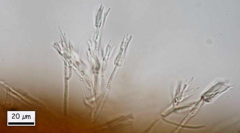
Miguel ûngel Ribes,
12-03-2013 11:27
Re : Hymenobolus agaves anamorph
Here there are.
Hans-Otto Baral,
12-03-2013 22:56
Re : Hymenobolus agaves anamorph
Hi Miguel
Walter Gams answered me that this is ô clearly aô Clonostachys, probably Clonostachysô solani (Harting) Schroers & W. Gams, which is quite common, often fungicolous, and the anamorph of a Bionectria. So certainly not belonging to Hymenobolus.
Zotto
Walter Gams answered me that this is ô clearly aô Clonostachys, probably Clonostachysô solani (Harting) Schroers & W. Gams, which is quite common, often fungicolous, and the anamorph of a Bionectria. So certainly not belonging to Hymenobolus.
Zotto
Miguel ûngel Ribes,
13-03-2013 00:15
Re : Hymenobolus agaves anamorph
Hi Zotto, Superb.
Thank you again to resolve this puzzle.
See you.
Thank you again to resolve this puzzle.
See you.