
05-06-2026 11:02
Thomas Læssøehttps://svampe.databasen.org/observations/10596691
07-06-2026 15:10
 William Slosse
Hello everyone,On 05-06-26, I found following asco
07-06-2026 12:43
Steve ClementsBojour. This was a strange find on a stick on my
07-06-2026 12:09
 François Freléchoux
Bonjour, Voici une brève description de ce qui m
12-07-2015 00:05
 Nedim Jukic
This one from the same locality as the previous on
06-06-2026 17:44
Steve ClementsBonjour, This disco was on planed wood 3 x 1.5 cm
14-08-2016 23:15
 Alex Akulov
Dear friendsCan you help me to find the descriptio

04-06-2026 11:36
Gernot FriebesHi,found on Vaccinium myrtillus.Asci: IKI –, 8-s
05-06-2026 12:10
François Freléchoux
Capitotricha sp. sur Lonicea caerulea Caractères
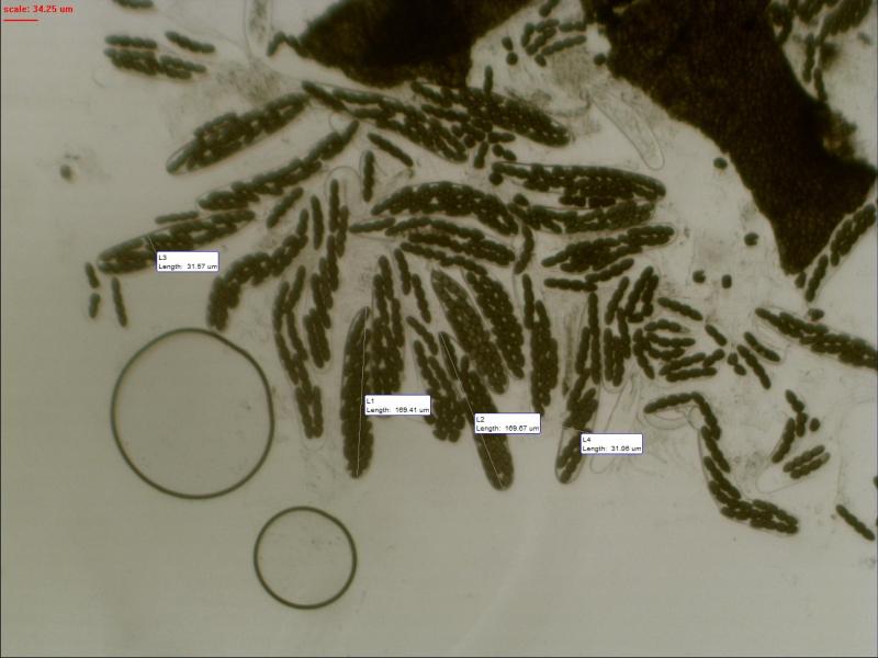
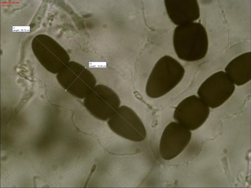
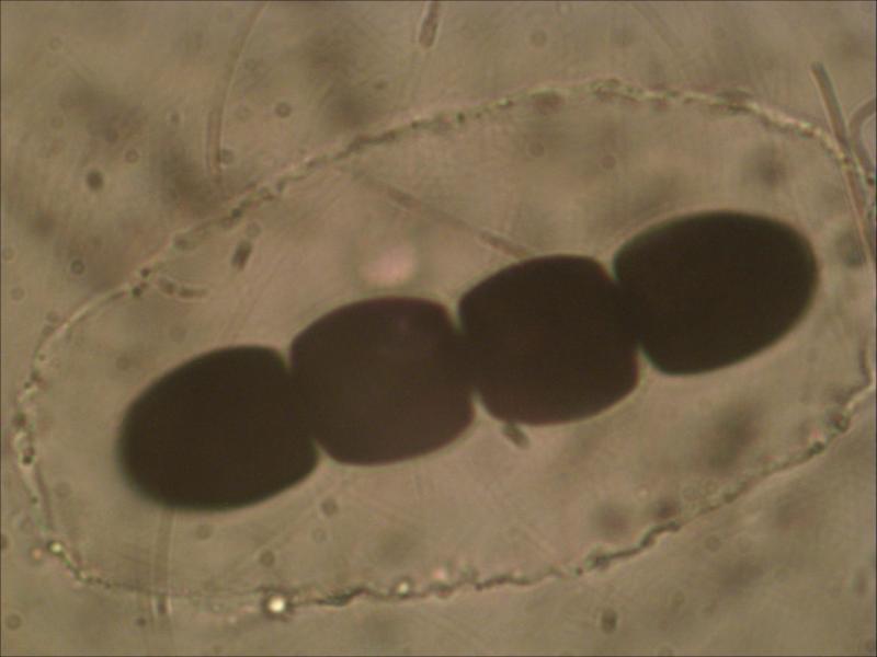
 Found on horse dung.
Asci: 169.41-169.67x31.06-31.57 um
Spore: 50.73x10.20-11.26 um
De second cell slightly smaller than the third one 12.14 over 12.41 um
Germ slit: Parallel to slightly oblique whereby they bent near the septum


yes, this should be S. megalospora, compare with my finding from Austria last year:
Sporormiella megalospora
regards,
björn

Sporesize and germslit conforms better with Sporormiella capybarae.
The spores of Sporormiella grandispora have a parallel germslit and slightly larger spores.
And Sporormiella megalospora has much larger spores.
Norbert
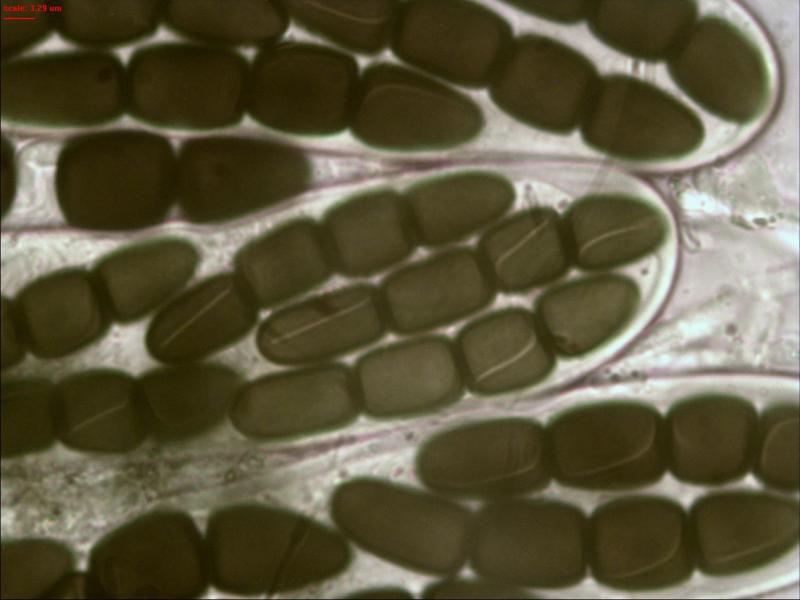
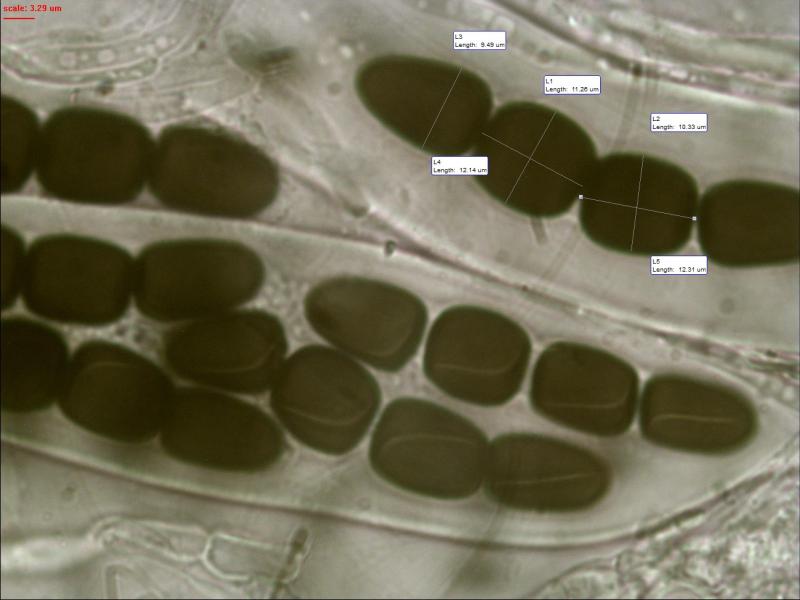
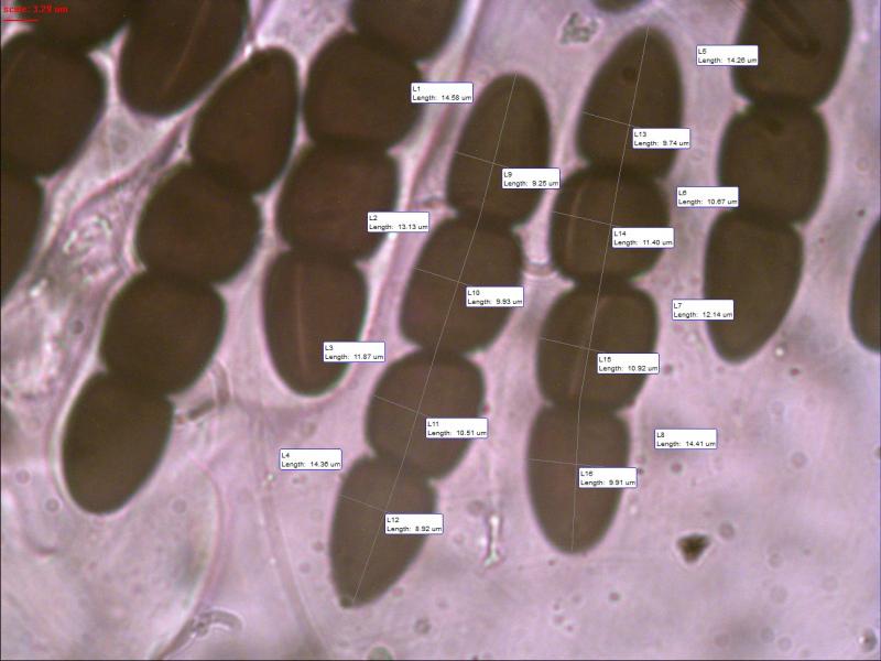
Hereby I send you a photo with measurements of each cell Norbert. Maybe this is a better confirmation.
Upper cell conical:
13.74x9.69; 13.08x9.17; 15.31x10.20 (extreme size)
Second cell barrel shaped:
10.55x10.40; 10.75x10.66; 13.16x11.81 (extreme size)
Third cell barrel shaped:
12.49x10.01; 12.46x9.67
Basel cell cylindrical rounded top:
13.99x9.12
Hopefully I can find more photo's of this one.
Do the germ slits also curve near the septum as they do with S. Grandispora Norbert?
In the article of Ahmed & Cain on page 442 about Sporormiella they also say for S. Grandispora that germ slits are usually parallel, occasionally slightly oblique, usually curved next to septum. Cells almost equal in size (in this case they ar not).


In the first photo, where we perceive the asci, they seem to have it as the base ends abruptly. Joop you can get a clearer picture? If this comfirme we would depart from S.capybarae which has asci gradually ending a long walk, and instead seek to S.intermedia.
Michel.
Michel


Michel.
Sorry, but in the moment I don't have enough time for an answer.
I'll write later.
Norbert
Is the shape of the gelatinous sheath a factor for determination?
They often differ in size and form, especially at the septa but they also can be fully cicular I will look up some photos to explain what I mean.
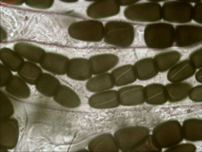
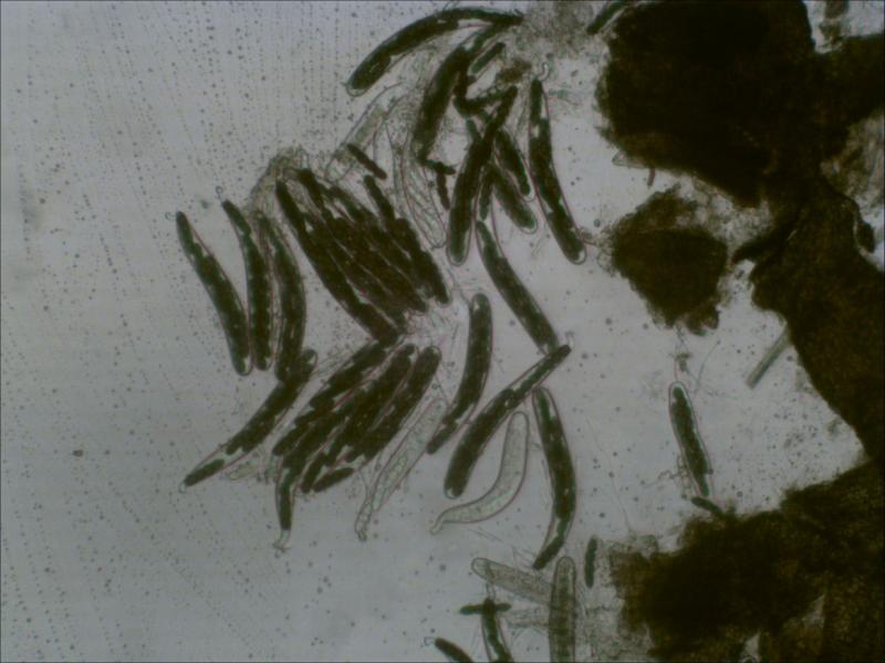
I also include a plhoto where you can see cells of sporormiella opening at the germ slit.


Michel.
Merci Michel
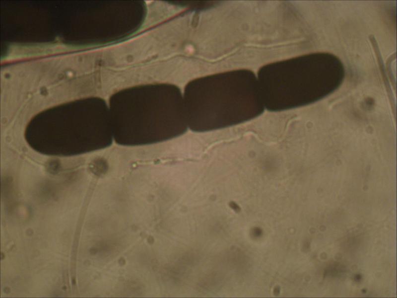
The other serie does not have short stalks for the asci I am still looking for better photo's that match.
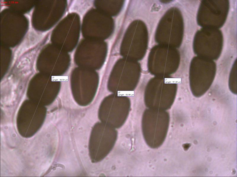
In the first serie the upper cell is conical, the second barrel shaped the third and also the basal end cell cylindrical with a rounded end for the latter.