

19-05-2026 12:55
 Hardware Tony
After checking Gminder and Otto's library I cannot
19-05-2026 10:27
Patrice TANCHAUDBonjour, récolte récente sur terre retournée i
18-05-2026 12:43
Sylvie Le GoffBonjour à tousPuis je avoir votre aide sur ce que
19-05-2026 14:56
Åge OterhalsI found this white cushion-formed ascomycete on ro

18-05-2026 10:13
 Lieve Deceuninck
Dear forum members,I identified this as the teleom

17-05-2026 19:05
Thomas FlammerI have found this tiny 200 ym cup shaped apothecia
17-05-2026 16:41
Margot en Geert VullingsWe found this Lachnum on an old Rubus stem.Fruitbo
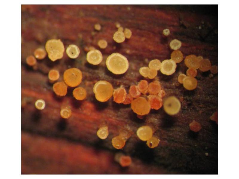
 Dear friends,
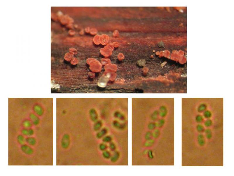
Today my wife find a Discomycete sample, which looks interesting. Its apothecia are formed on thin twigs of unknown deciduous tree with signs of discoloration in red. Young apothecia are yellow, but soon stay red as the wood .
Asci are 8 - sporous, very small, in average of 14 x 4.2 micrometers. Asci are both monostichous and distichous, but more frequently 6 spores at the top of ascus are placed in two rows, and the two spores near the ascus leg are placed in one row. The size of ascospores: 3.1-3.3 x 1.7-2.2 micrometer.
Please, tell me what is this species and whether it is the cause of red staining of wood or vice versa it absorbs red pigment from the wood.
Thanks in advance ,
Alex

it might be a Hyphodiscus, but to clarify you should lok for marginal hairs, and whether it has a gelatinized excipulum.
H. hymenipohilus is known to stain the wood in red by its anamorph.
Do you have a photo of an ascus? is it amyloid?
Zotto
Thanks for your advice! In the Lugol reagent apical apparatus of asci colored in blue. Most of the asci in my sample are immature, but as a whole the morphology of asci, spores and marginal hairs are very similar to those shown on the site: http://asco-sonneberg.de/pages/gallery/hyphodiscus-hymeniophilus-091227-mcol-0115551.php?group_id=15511&position=2
The legs of asci are enough long and their overall size, even in an immature state is 37 x 4.3 micrometer.
Probably I'm dealing with a young specimen of Hyphodiscus hymeniophilus.
Alex