
23-05-2026 18:57
Sylvie Le GoffBonjour à tousRécolté sur une branchette de Sal
22-05-2026 14:44
 Lothar Krieglsteiner
in unripe condition citrine yellow, then soon fadi
23-05-2026 11:44
 Charles Grapinet
Hello, I am having trouble identifying this copro
22-05-2026 21:35
Steve ClementsBonjour, I expected this find on old wood on our
22-05-2026 18:12
Lothar Krieglsteiner
... in moist chamber from Portugal.As the fungus s
22-05-2026 20:08
Ethan CrensonHello all, Yesterday in NYC I was visiting an e
11-01-2022 16:36
 Jason Karakehian
Hi does anyone have a digital copy of Raitviir A (
20-05-2026 17:47
Margot en Geert VullingsWe found this Mollisia on dead Juncus stems mown l
22-05-2026 14:47
Gernot FriebesHi,superficial ascomata collected on bark of a liv
Hello!
I thought that I sent this... This one was maybe ten meters from jelly fungi on Hypogymnia tubulosa. This was on Phaeophyscia or Physcia sp.
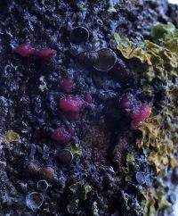

if you mean this red objects, check Illosporiopsis christiansenii, a parasite on lichen.
regards
Martin
It looks same. Do you know how common it is in North-Europe?
regards
Martin

The photo is a little small and perhaps we can also think at Marchandiomyces corallinus.
Microscopy would be useful here again.
Alain
we had a thread in the German microscopy forum:
http://www.mikroskopie-forum.de/index.php?topic=18152.15
there I posted the microscopical features of both taxa.
(the thread starts here: http://www.mikroskopie-forum.de/index.php?topic=18152.0)
Martin
Hi Martin,
You have very interesting discussion in Gemany :)
Photographies are very good.
Alain