05-06-2026 12:10
 François Freléchoux
Capitotricha sp. sur Lonicea caerulea Caractères
05-06-2026 11:02
Thomas Læssøehttps://svampe.databasen.org/observations/10596691

04-06-2026 11:36
Gernot FriebesHi,found on Vaccinium myrtillus.Asci: IKI –, 8-s
19-05-2026 10:27
Patrice TANCHAUDBonjour, récolte récente sur terre retournée i
04-06-2026 18:39
Gernot FriebesHi,I collected this species in two different locat
22-05-2026 13:29
Gernot FriebesHi,I am curious to hear your opinion on this mater
04-06-2026 10:50
François Freléchoux
Bonjour, J'ai trouvé hier un petit asco observé
 Hello,
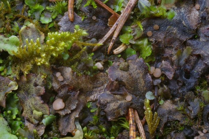
I would welcome your advice on this Bryoscyphus.
It was collected in Slovakia, Mala Fatra NP, in a gorge, on old thalli of Preissia quadrata, 10 May 2015.
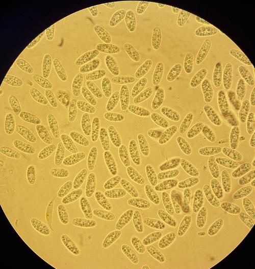
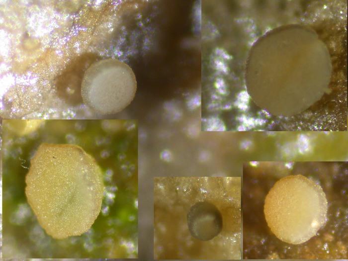
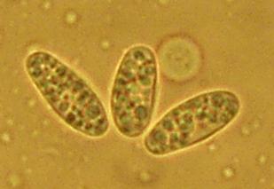
Apothecia grew in small groups, were slightly convex or plane, +- rounded from an upper view, pale brown, 0.5-2.5 mm in diameter, shortly stipitate.
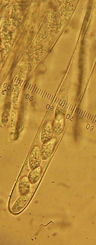
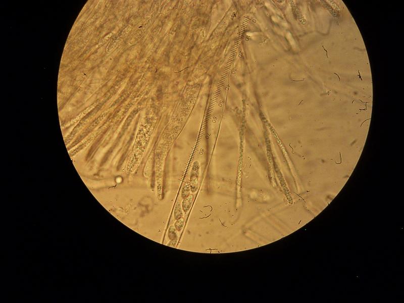
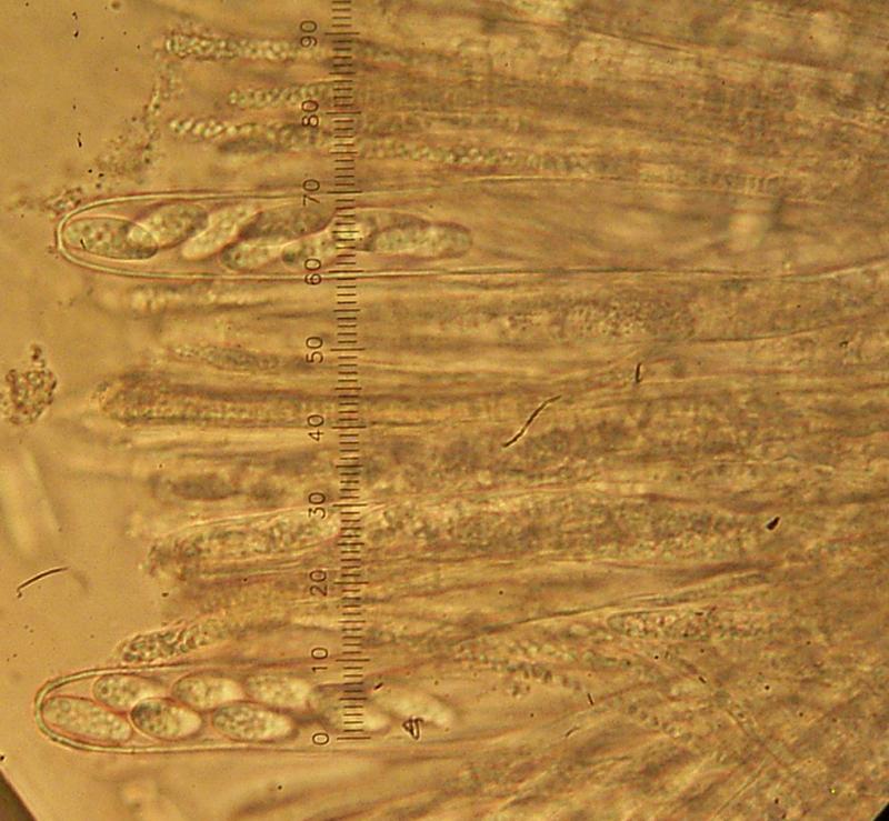
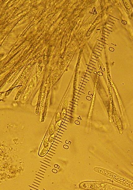
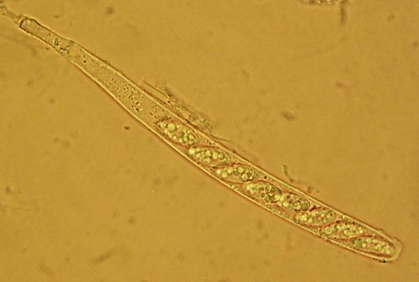
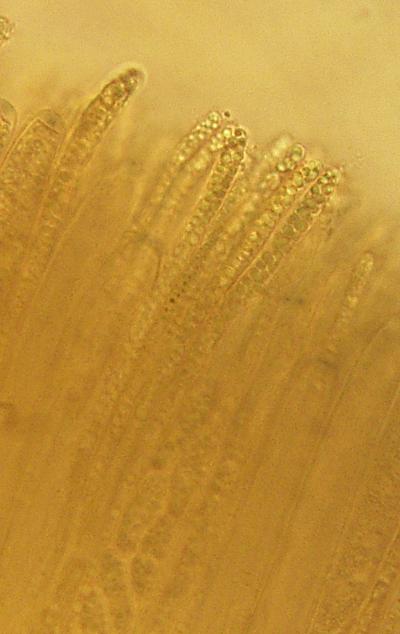
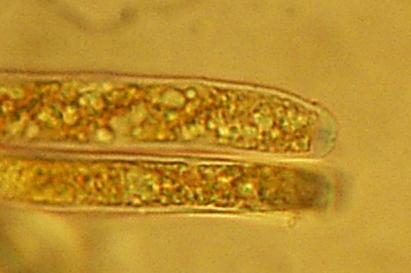
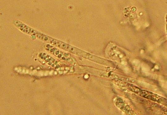
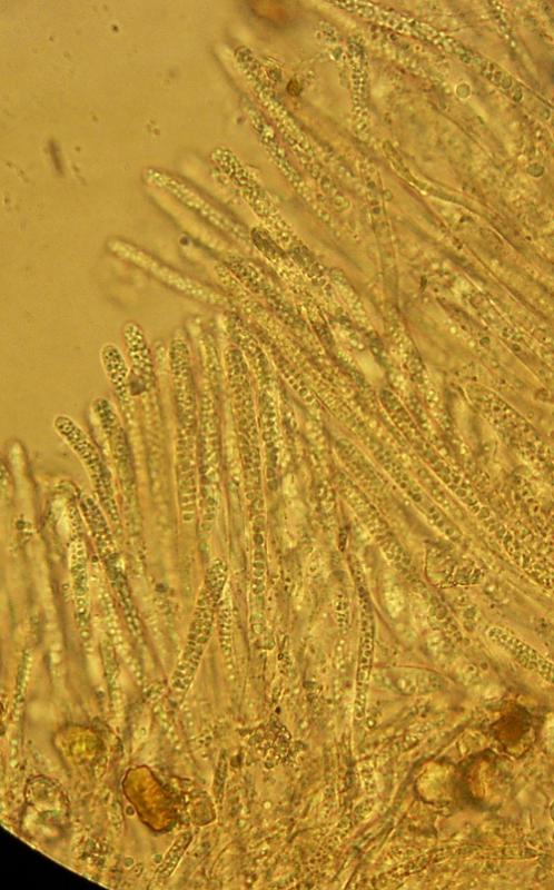
Asci cylindrical, inoperculate, octosporic, uniseriate or biseriate, IKI+, 130-160 x 9-12 micrometers.
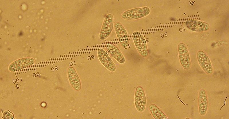
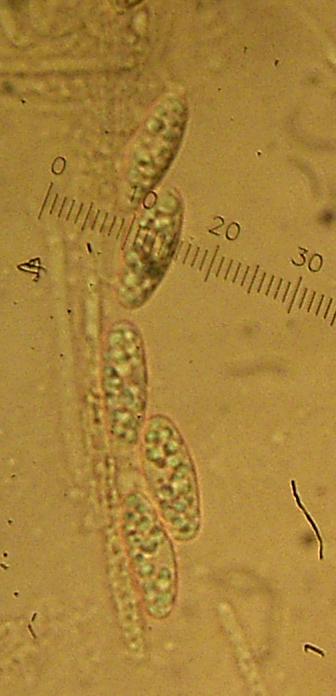
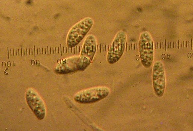
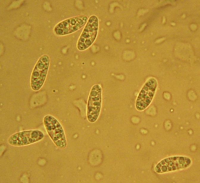
Spores unequilateral, smooth, with many small droplets, 12.5-15(17) x (4.5)5-5.5(6) micrometers.
Paraphyses filled with small droplets, hyaline or pale brown, septate, branched, with an obtuse apex, 3-3.5 micrometers broad, straight.
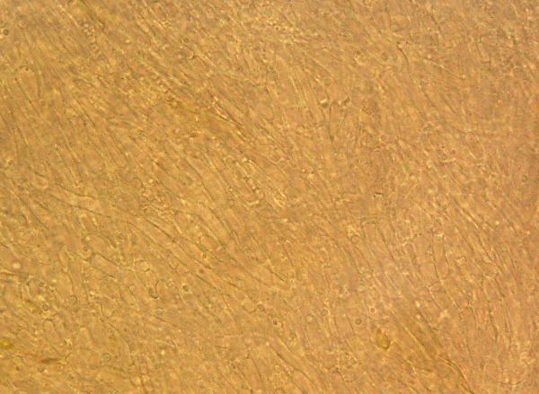
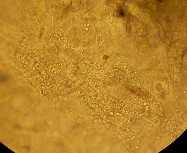
Med. excipulum consisting of cylindrical thin-walled cells (2.5-4 micrometers broad).
Ectal excipulum combination of textura angularis and text. prismatica. Hair-like projections not observed.
No combination of characters in the key in the article by Verkley et al. 1997 fits perfectly. Preissia (member of Marchantiaceae) is not mentioned as a substrate in any of the species in the key. B. dicrani and B. turbinatus have totally different hosts, so I´m considering B. atromarginatus, B. marchantiae and B. conocephali. The width of spores is between B. atromarginatus and B. marchantiae, asci are too long for B. atromarginatus, spore shape wrong for B. marchantiae. For B. conocephali, the hairs are missing.
Zuzana
Je pense qu'il s'agit de B. atromarginatus Verkley, van Aa & G. de Cock. Voir ma publication en dossier attaché.
Amicalement
René
DOUGOUD, R. (2003) - Bryoscyphus atromarginatus. - Mycol. Montenegrina VI: 7-11.
René

Dear Rene,
I would also like to receive the file. Thanks in advance,
Yours, Lothar Krieglsteiner

Bonjour Zuzana, René et tous,
Belle récolte et belles photos.
I did not recognize the spores of a last year collection I called B. atromarginatus (on Lunularia cruciata). As in René's paper (which I also used for identification) the spore shape is different , along with guttulation . Spore width of my collection (MH 120114) never exceed 4.5 µm and there always is an empty area in the spore guttuation.
Asci never exceed 120 (130) µm
I do not know B. marchantiae.
I attach pics .Amitiés Michel
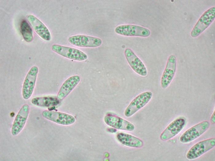

this is actually striking how the nucleus is in the lower part of the spore in your fungus. In all my available images I see the empty region +/- in the middle part.
Spore width seems variable, I have 3.5-4.5 up to rarely 5-5.5 µm.
Of course, I am not sure if all is the same, but i would at first sight think that yours belongs here.
Zotto
In my collection the spores often have an empty zone too, but rather small. I add some new photos.
Comparing my data with those in René´s article, I can see few differences - the substrate, somewhat different size of spores and asci (I measured everything fresh, in water). If I understand the French text well, René´s paraphyses were simple (mine are sometimes branched, see the photo with two lateral branches). I also never observed septa in the spores.
But these differences are probably not very important.
Best regards, Zuzana
Septation occurs only in overmature spores in this group, so it depends on the ripeness of the apos.
Zotto