
18-03-2026 17:22
Katarina PastircakovaHi there,I'm looking for the following literature:
27-11-2025 15:41
Thomas LæssøeSpores brownish, typically 4-celled; 26.8 x 2.4;
18-03-2026 13:09
 Khomenko Igor
I recently examined Celtis occidentalis branches
18-03-2026 11:52
Thomas Læssøehttps://svampe.databasen.org/observations/10493688
11-03-2026 17:36
 Michel Hairaud
Bonjour, Je cherche des indices pour cette réc
17-03-2026 10:40
 Martine Vandeplanque
Bonjour à tous.Chaque année en mars ou avril, il
17-03-2026 19:41
 Bernard CLESSE
Bonsoir à toutes et tous,Pourriez-vous m'aider à
12-03-2026 19:44
 Enrique Rubio
Hi to everybody.Can you give me any suggestions ab
17-03-2026 10:09
 François Freléchoux
Bonjour, Voici la description rapide d'un petit d
05-03-2026 10:07
Hulda Caroline HolteHello, I found and collected this species growing
 Greetings AscoFrance!
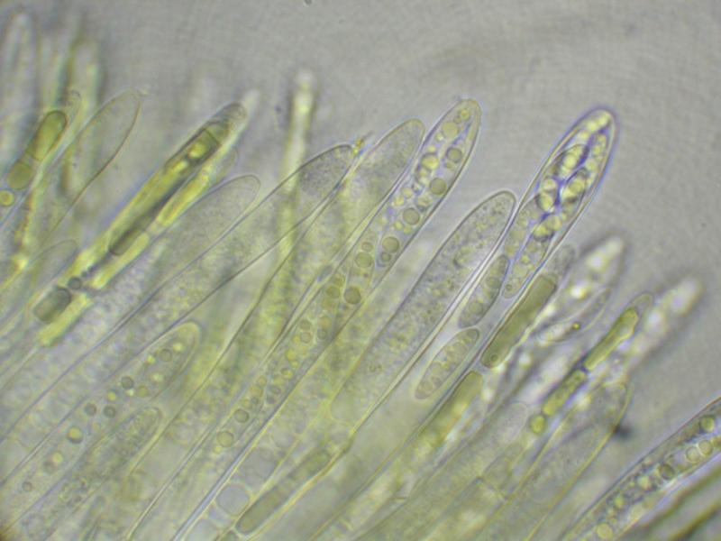
Here is a curious discomycete, field IDed to Ionomidotis by Paula DeSanto on the recent Peck Foray in Watkins Glen, New York. This particular find is from a mixed, predominantly hardwood forest within the Meads Creek State Forest. When I got a look at the dried material and field photos, I saw enough resemblance to my own Ionomidotis collection from North Carolina (http://mushroomobserver.org/174774) to consider the possibility, but upon preparing the material for microscopy we noticed that KOH extractable pigments (3% solution) were conspicuously absent. Can it still be Ionomidotis without this reaction? Perhaps this is a member of some other genus in the Encoelioideae?
Ascus tips inamyloid, despite appearing somewhat bluish in the micrographs. No paraphyses observed.
Spores:
9.5-14×=2.5-4.5?m (x=12.25×3.325?m, Q= 2.44-5.2?m, Qm=3.781?m, m=20, s=1)
13.5 x 3 ; 4.5
13 x 4 ; 3.25
13 x 2.5 ; 5.2
12.5 x 3.5 ; 3.57
14 x 4 ; 3.5
13 x 3 ; 4.33
9.5 x 3 ; 3.17
12.5 x 4 ; 3.13
13 x 3.5 ; 3.71
11 x 4.5 ; 2.44
13.5 x 3 ; 4.5
13.5 x 3 ; 4.5
10.5 x 4 ; 2.65
12 x 3 ; 4
12.5 x 2.5 ; 5
12.5 x 3 ; 4.17
9.5 x 3 ; 3.17
14 x 4 ; 3.5
11.5 x 3 ; 3.83
10.5 x 3 ; 3.5
Many thanks!
-Danny N.
PS: The images are all apparently too large for the site :( Please find them on Mushroom Observer here: http://mushroomobserver.org/218595

I am reminded of a Chlorencoelia, but the two species for which I have images, C. versiformis and C. torta) have distinctly amyloid asci. The spores would fit.
I am sure that the paraphyses would be seen when squashing the hymenium. If you had pictures from fresh material the genus Chlorencoelia would show a striking feature in the paraphyses (vacuolar bodies, see attach).
Zotto
Also, I believe the fact that the bottle of Melzer's used was labelled "Melzer's Replacement" may have something to do with the lack of observed blueing. Will use a more reliable reagent for the second set of micrographs.
These vacuolar bodies are a useful character at the family level. They are rather typical for the family Cenangiaceae as we now circumscribe it, but absent from the Cordieritidaceae which inbclude many ionomidotic species.
many thanks!