

23-03-2026 20:16
 Miguel Ángel Ribes
Good eveningI'm unable to identify this Coprotus o
21-03-2026 15:13
Lepista ZacariasHello everyone, Does any one know of any literatu
20-10-2017 09:23
Garcia SusanaEste otro crecía en el mismo trocito de madera qu
20-03-2026 16:16
 Edvin Johannesen
These 0.5 mm diam. acervuli were breaking through
19-03-2026 19:34
 Filip Fuljer
Hello everyone,a few days ago I collected this str
19-03-2026 18:25
 William Slosse
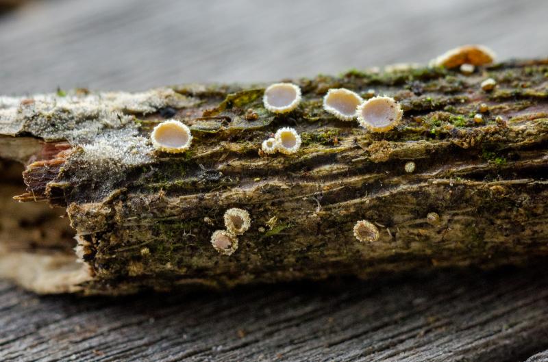
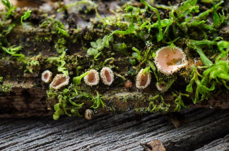
Good evening everyone, On 18/03/26 I found a few
 http://mushroomobserver.org/230585
Trichopeziza?
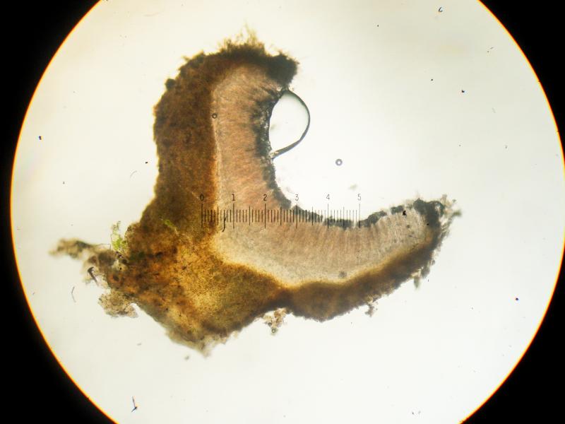
Only these poor macro and micro images to go on. No magnifications, no certainty on scope calibration or mounting mediums.
Any and all help greatly appreciated.
-myxomop

I don`t know if it is help and you are right: there is few information (and Costa Rica is far from Europe) - but I see some similarity of your fungus with Perrotia tricolor.
Surely Zotto will contradict.
Regards from Lothar

this looks like something interesting for me. Does this collection exist or only the photos?
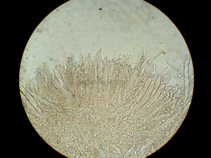
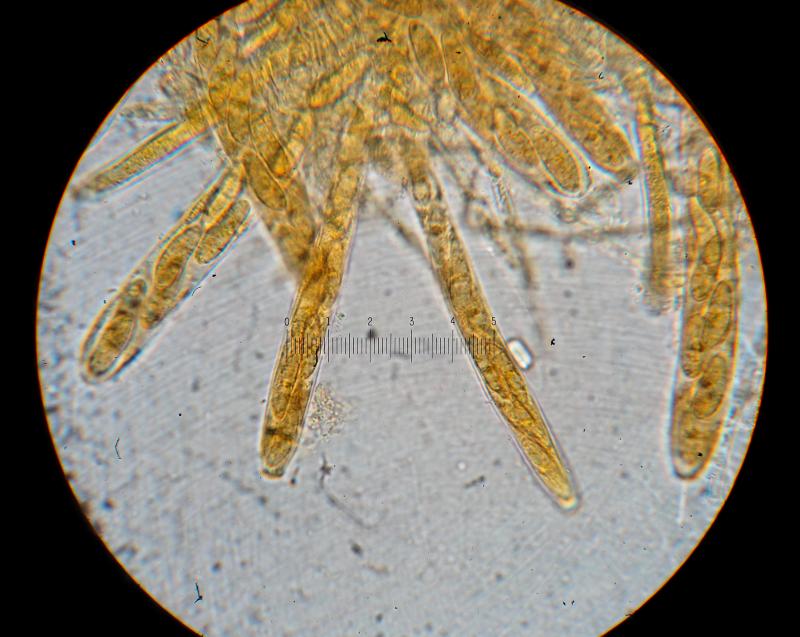
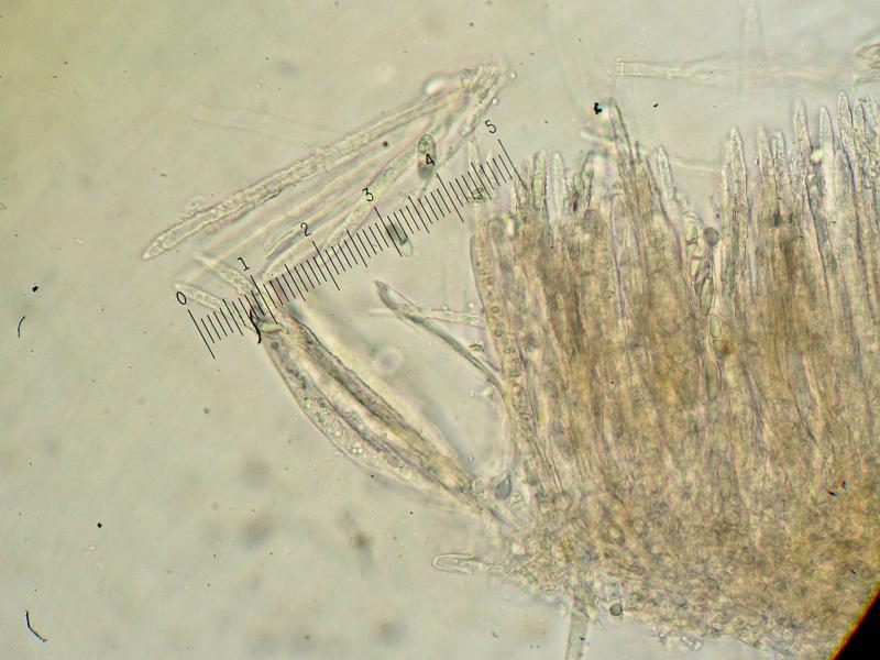
The paraphyses have a rough surface by some exudate which is known in a few members of Trichopezizoideae, but not from typical species of Trichopeziza. For instance I saw this in a collection of Trichopezizella barbata and in a number of apparently undescribed taxa. Maybe these deserve a genus of their own, and we are presently trying to find out about it by sequencin a number of species.
Your collection could also be compared with the genus Erioscyphella in which a lot of tropical species belong, such as E. abnormis but I don't see a species with such spores. You don't have a photo of the hairs?
Zotto
Proliferodiscus tricolor is similar, yes, but has cylindrical non-protruding paraphyses without these deposits on them.
I'm afraid these are the only images. Thank you for your comments and suggestions, despite the lack of more information.
Best,
-Danny
Sessile, up to 2mm in diam.
Asci ~115-150µm x 10-15µm, 8-spored, inoperculate, inamyloid. Paraphyses ~175µm x 5µm. Hairs ?µm x 4-5µm, multi-septate.
Spore Measurements:
15-22.5µm x 5-7.5µm (x=19.66µm x 6.6µm, m=20, s=1)
22.5x7.5
21.5x7.5
22.5x7.5
21.5x7.5
21.5x7.5
17.5x7.5
22.5x7.5
20x6.5
18.5x6.5
18.5x6.5
15x5
20x6.5
18.5x5
17.5x5.5
17.5x5
17.5x5.5
20.5x7.5
20x7.5
19x5.5
20.5x7
I'm guessing I did not get hair length measurements because they were enormously long.
Does any of this get us closer to an ID?
My continued thanks,
-Danny
If really inamyloid, your fungus indeed comes close to my T. perrotioides, except for the much larger spores. A pity that it is not preserved.
I can't find a reference to T. perrotioides. Is this a published name? Is it a Trichopeziza or Trichopezizella?
Also, I believe at least some of the micrographs represent fresh, living material, though I cannot 100% confirm this.
If some spores or paraphyses were alive the contents are not very good to see. Good is to have free spores that were forcibly ejected from the living asci, then you have a very constant and taxonomically significant arrangement of guttules.