

11-04-2026 10:19
 Michel Hairaud
Chers amis d'Ascofrance , voici une très bonne no
11-04-2026 10:10
Michel Hairaud
Dear Ascofrance members, here is some very good ne
10-04-2026 23:22
Gernot FriebesHi,ascospores are 1- to 3-septate, approximately
10-04-2026 15:51
 William Slosse
Hello everyone, On 08/04/26, I found a growth sit
09-04-2026 15:25
Jac GelderblomOn bare soil between mosses Ifound an asco I deter
09-04-2026 13:55
Thomas Læssøehttps://svampe.databasen.org/observations/10589176
09-04-2026 10:12
Thomas Læssøehttps://svampe.databasen.org/observations/10587061
08-04-2026 20:33
 Vasileios Kaounas
Found 07-04-26, in Abies cephalonica. Diameter 1,
08-04-2026 10:39
FRANCIS FOUCHIERBonjour , je recherche en pdf cet article: KORF R
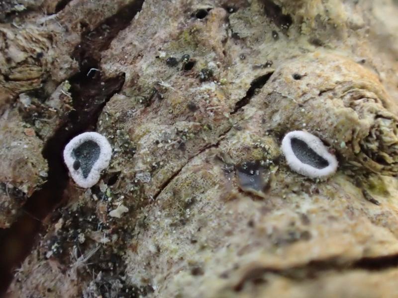
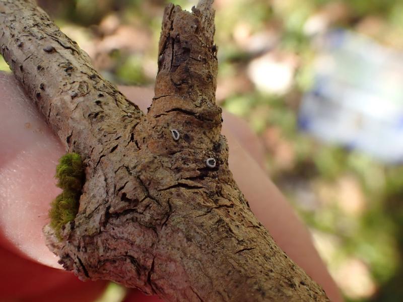
Discomycete with muriform(?) spores on Sambucus racemosa
Edvin Johannesen,
06-10-2020 14:04

Hi!
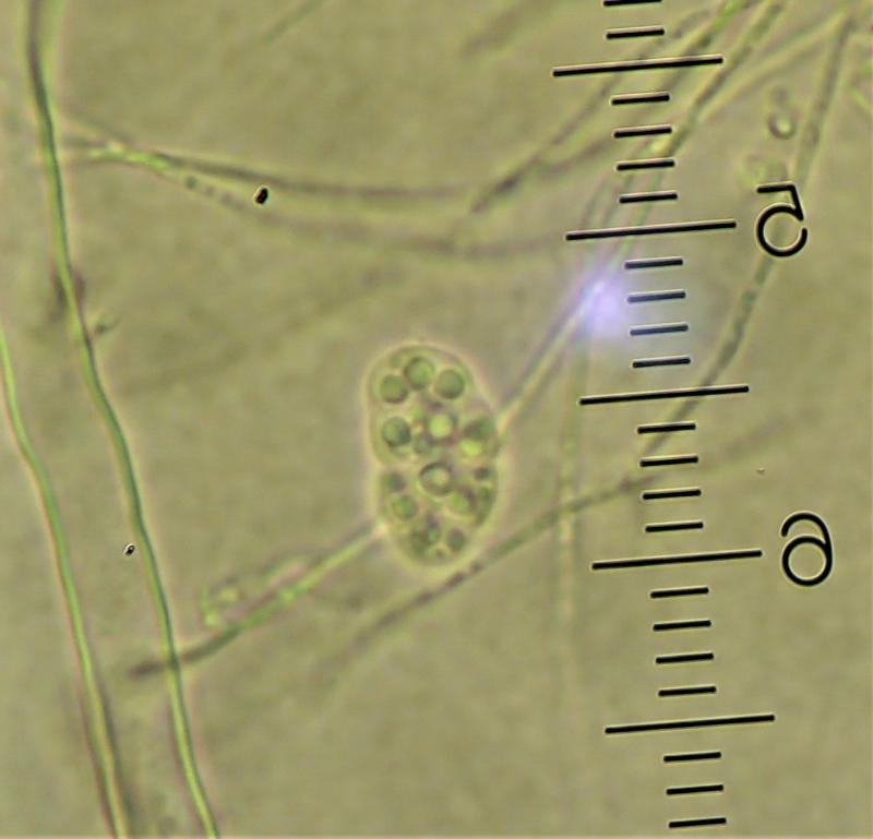
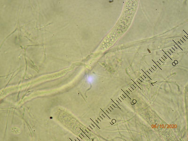
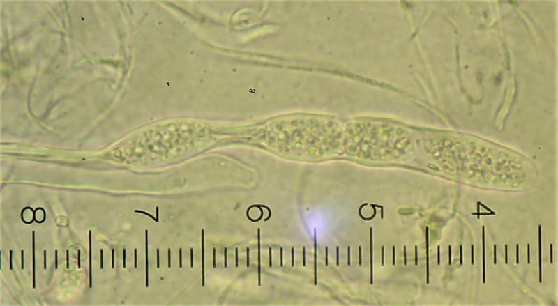
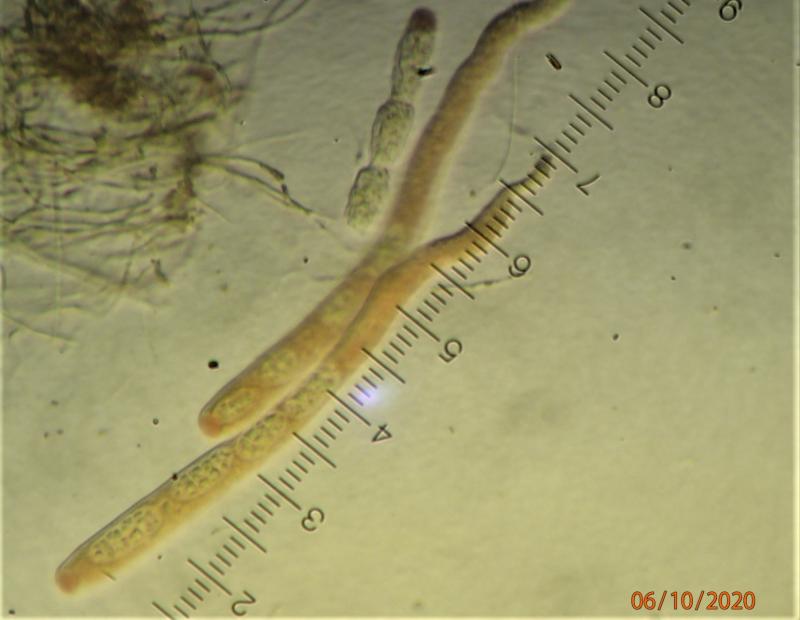
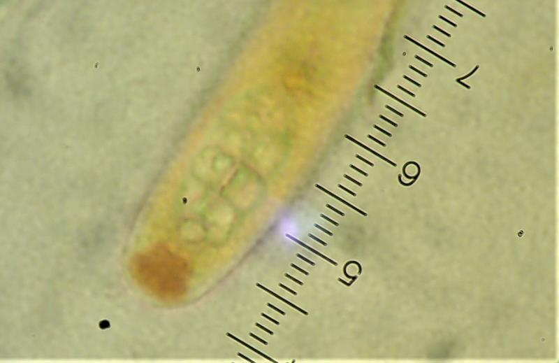
I am struggling with this tiny disco with a grey hymenium and a white, downy rim. I have only two apothecia, so not much to play with. Asci measure approx. 200 x 10 microns (in water) - shorter (ca. 150) in Lugol. Spores ca. 17 x 9-10 microns. They are either muriform or multiguttulate - it's hard to tell - and hyaline. The entire ascus "cap"is reddish-brown in Lugol (no pre-treatment with KOH). Paraphyses filiform, ca. 1 micron thick. Asci appear to be 4-spored or else 4 spores are consistently aborted.
Suggestions appreciated. Thanks.
Edvin Johannesen,
06-10-2020 14:05
Re : Discomycete with muriform(?) spores on Sambucus racemosa
Microscopic details in water
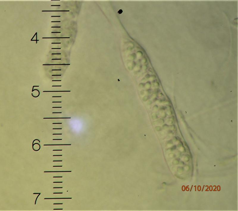
Edvin Johannesen,
06-10-2020 14:06
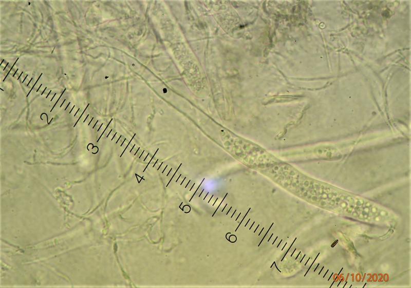
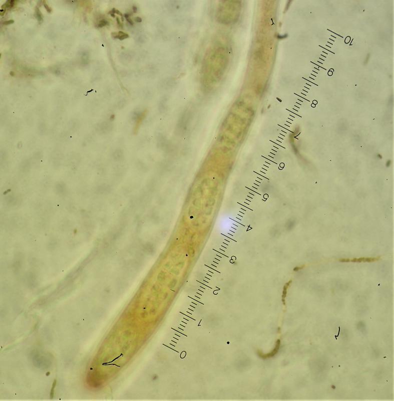
Re : Discomycete with muriform(?) spores on Sambucus racemosa
In Lugol
Martin Bemmann,
06-10-2020 15:44

Re : Discomycete with muriform(?) spores on Sambucus racemosa
Hi Edvin,
perhaps a lichen. There seem to be enough algae visible on the macrophoto.
regards
Martin
Hans-Otto Baral,
06-10-2020 17:06

Re : Discomycete with muriform(?) spores on Sambucus racemosa
I cannot see pic 3 and on the other I do not see algae. The red IKI reaction of the apical ring is interesting. I suggest to test also IKI after KOH, whether the ascus wall also reacts.
The thin paraphyses seem strongly branched.
Cryptodiscus muriformis resembles somewhat your specimen, including the 4-spored asci, but the spores are much larger and the apos orange. Here the entire ascus wall reacts, but also a distinct apical ring.
Important would be a section to study the margin, and perhaps also the presence of algae.
The large oil drops mask the cell walls. If you mount in MLZ or CB, or CR, you will better see the walls.
Edvin Johannesen,
06-10-2020 17:38
Re : Discomycete with muriform(?) spores on Sambucus racemosa
Thanks to both for your comments. It turns out that this one has been collected several times along the coast of Norway. A paper is in progress. I think one of the author will comment here. Sambucus is a new host/substrate.