05-06-2026 11:02
Thomas Læssøehttps://svampe.databasen.org/observations/10596691
07-06-2026 15:10
 William Slosse
Hello everyone,On 05-06-26, I found following asco
07-06-2026 12:43
Steve ClementsBojour. This was a strange find on a stick on my
07-06-2026 12:09
 François Freléchoux
Bonjour, Voici une brève description de ce qui m
12-07-2015 00:05
 Nedim Jukic
This one from the same locality as the previous on
06-06-2026 17:44
Steve ClementsBonjour, This disco was on planed wood 3 x 1.5 cm
14-08-2016 23:15
 Alex Akulov
Dear friendsCan you help me to find the descriptio

04-06-2026 11:36
Gernot FriebesHi,found on Vaccinium myrtillus.Asci: IKI –, 8-s
05-06-2026 12:10
François Freléchoux
Capitotricha sp. sur Lonicea caerulea Caractères
Sporormiella longisporopsis
Joop van der Lee,
14-10-2020 10:48
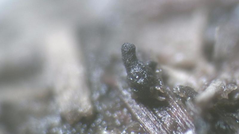
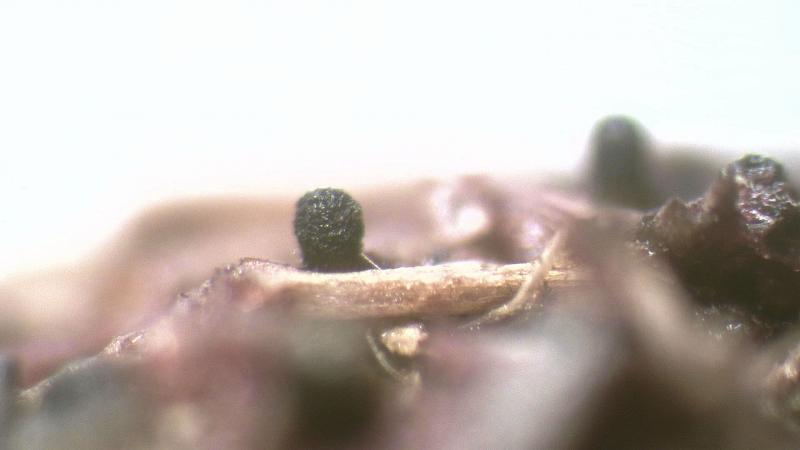
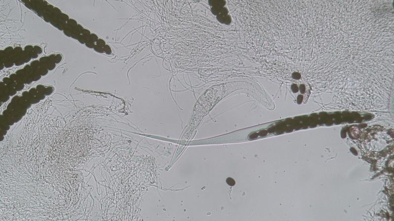
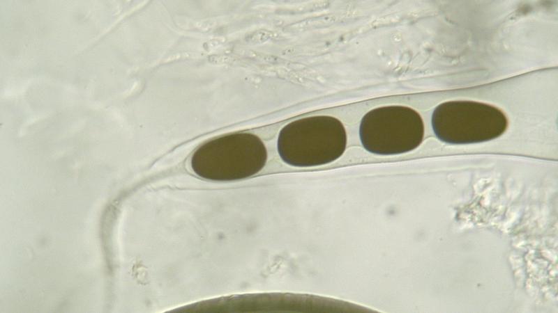
 Found on rabbit pellets and to my knowledge the first time in Holland.
Perithecia immersed with only the top and a small part opf the hyaline neck emersed (photo-1), 540x385 um, long hyaline neck with a dark brown top, total length 678x161 um of which 458 is hyaline and a top of 220x181 um (photo-2).
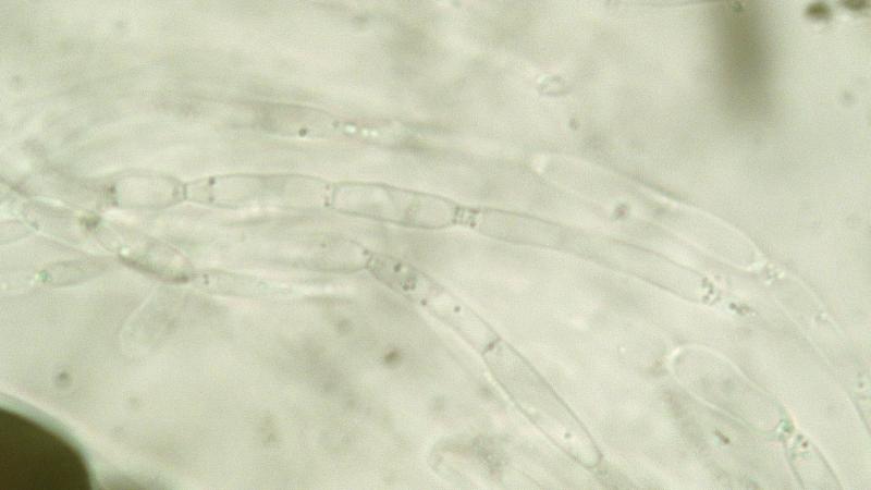
Paraphyses: Long septated hyaline cells 56.5-67x6.0-7.5 um measured between two consecutive deep constrictions (photo-3).
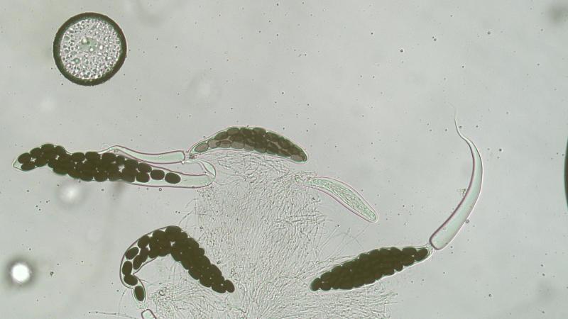
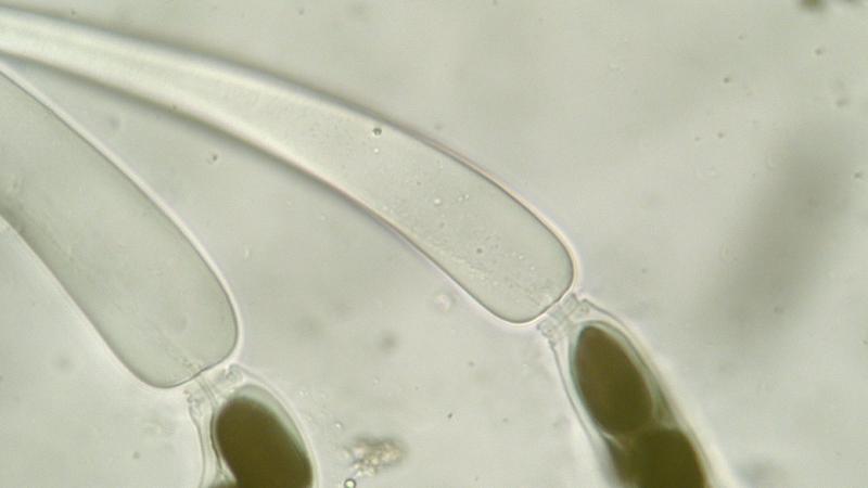
Asci: 8-spored, 215x50 um with mature spores and 205x41 um with immature spores (photo-4).
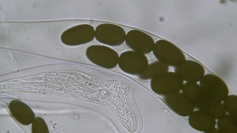
Asci can extend to 736x32 um with an extremely long tail (photo-5).
They also creates a secondary tail from the stolk connection downward enabling spores to enter (photo-6)
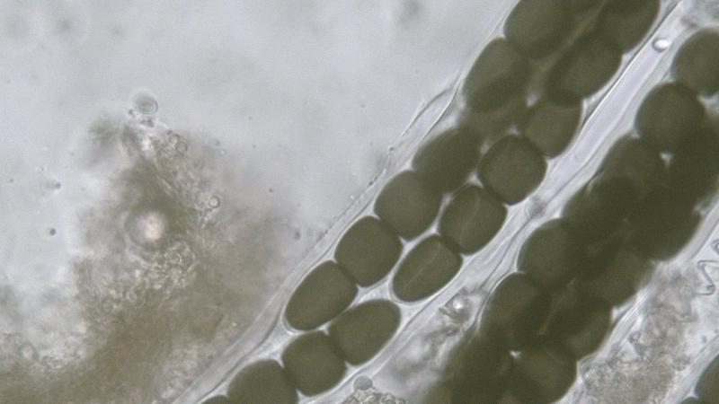
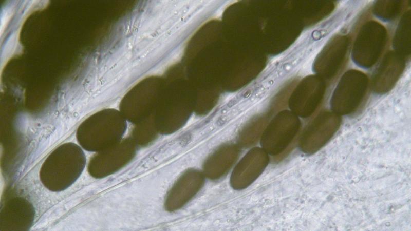
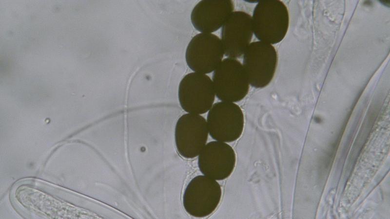
Spores: 4 selled, multi seriate, parallel germ slit spore 86x12 um (photo-7) and a solid gelatinous sheath 19 um wide spore 107.5x14.8 um (photo-8).
When cells separate inside the ascus they ar still connected by a gelatinous substance (photo-9)
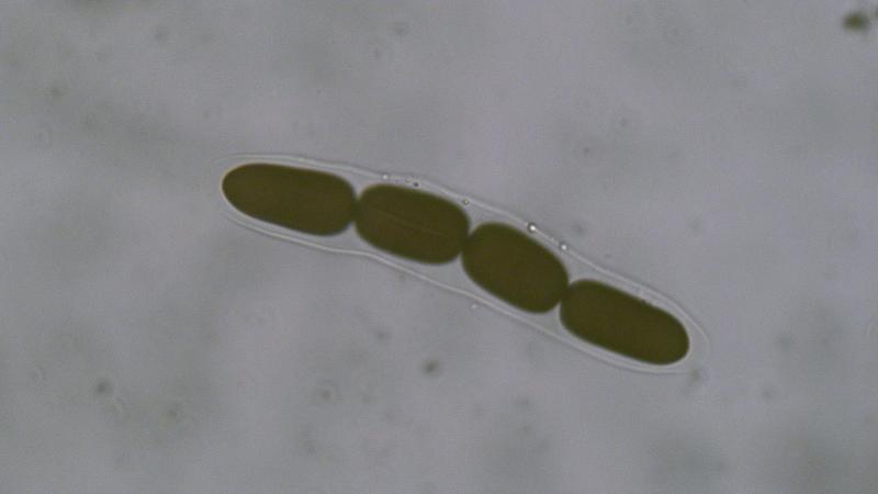
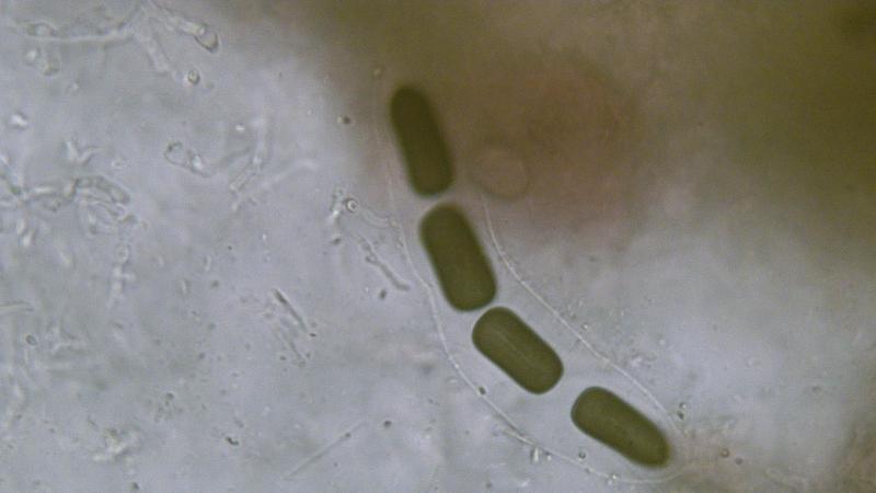
Spores measurements differ over a large range depending on the maturity of spores, roughly said its range is from 79-107.5x12-17.5 um.
Photo 10 103.5x15.8 um
Photo 11 79x17.5 um, oval
Photo 12 spore cells average 23 um except 2nd from basal which measures 21 um.
S. longisporopsis can be distinguished from S. longispora by the the difference in spore cell corners, for S. longisporopsis they are distinctly rounded and for S. longispora they are rectangular and S. longispora has a short neck.
When a small part of the hyaline neck is not sticking out of the dung it can easily be misteken for another species because of the broad neck resulting in a separation from the neck and the perithecia will than remain in the dung and never be found.
Joop
Yulia Lytvynenko,
18-10-2020 05:01

Re : Sporormiella longisporopsis
Hi, Joop.
I also have samples of this species on horse and goat dung.
Asci 8-spored, cylindrical-clavate, gradually tapered below into a stout, persistent stipe, 289–296 × 36–39 ?m.
Ascospores bi- or tri-seriate, 4-celled, 80,6–98,0 × 14,5–17,6 ?m, cylindrical, straight or slightly curved, yellowish brown when young to dark brown when mature; septa transverse, constrictions at septa broad and deep; segments easily separable, all cells are almost equal in length, roundish at the apex; germ slits parallel or oblique.
S. longisporopsis is probably a rare species in the world, as noted by some other researchers (Doveri 2004, Mungai et al. 2012).
Joop van der Lee,
18-10-2020 12:09
Re : Sporormiella longisporopsis
Hello Yulia,
I also found species that looked like S. longisporopsis with a shorter neck.
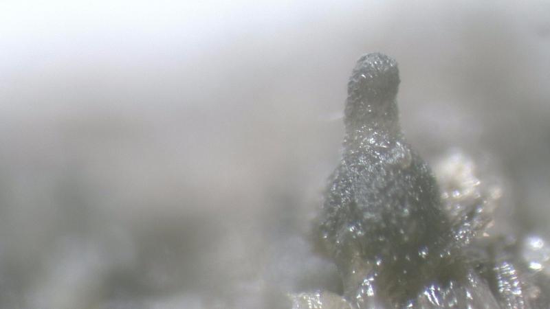
Under the microscope it turned out to be S. megalospora specifically due to the germ slit pattern being a good classifier and the shape of the ascospores barrel/bulbous shaped agains t oval although in the topic about S. longisporopsis spores appeared bulbous as well (photo-11) so maybe they are interconnected.
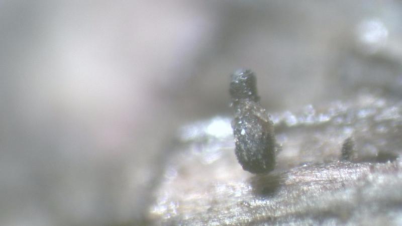
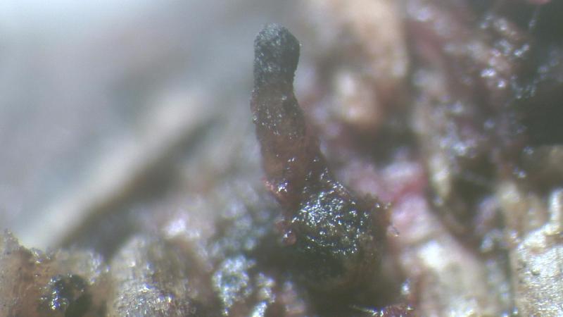
The last photo shows a S. longisporopsis having a longer neck.
I also noticed that often two species of the same family appear in a group in particular on rabbit pellets.
Schizothecium conicum with Schizothecium aloides
Schizothecium vesticola with Schizothecium tetraspora
Podospora setosa with Podospora curvicolla
Sporormiella longisporopsis with Sporormiella megalospora
By the way the current name for this species is Preussia longisporopsis
Joop
I also found species that looked like S. longisporopsis with a shorter neck.
Under the microscope it turned out to be S. megalospora specifically due to the germ slit pattern being a good classifier and the shape of the ascospores barrel/bulbous shaped agains t oval although in the topic about S. longisporopsis spores appeared bulbous as well (photo-11) so maybe they are interconnected.
The last photo shows a S. longisporopsis having a longer neck.
I also noticed that often two species of the same family appear in a group in particular on rabbit pellets.
Schizothecium conicum with Schizothecium aloides
Schizothecium vesticola with Schizothecium tetraspora
Podospora setosa with Podospora curvicolla
Sporormiella longisporopsis with Sporormiella megalospora
By the way the current name for this species is Preussia longisporopsis
Joop