21-03-2026 15:13
Lepista ZacariasHello everyone, Does any one know of any literatu
20-10-2017 09:23
Garcia SusanaEste otro crecía en el mismo trocito de madera qu
20-03-2026 16:16
 Edvin Johannesen
These 0.5 mm diam. acervuli were breaking through
19-03-2026 19:34
 Filip Fuljer
Hello everyone,a few days ago I collected this str
19-03-2026 18:25
 William Slosse
Good evening everyone, On 18/03/26 I found a few
17-03-2026 10:09
 François Freléchoux
Bonjour, Voici la description rapide d'un petit d
19-03-2026 17:50
 Enrique Rubio
Hi to everybodyThese thiny, blackish pseudothecia
Phyllocharis orbicularis
Roo Vandegrift,
28-09-2022 22:03
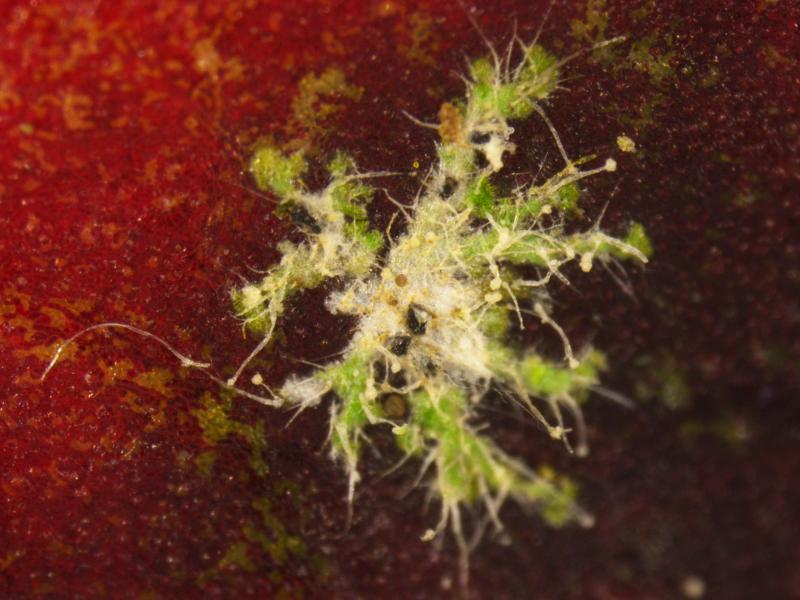
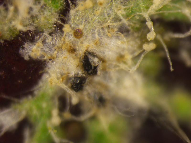
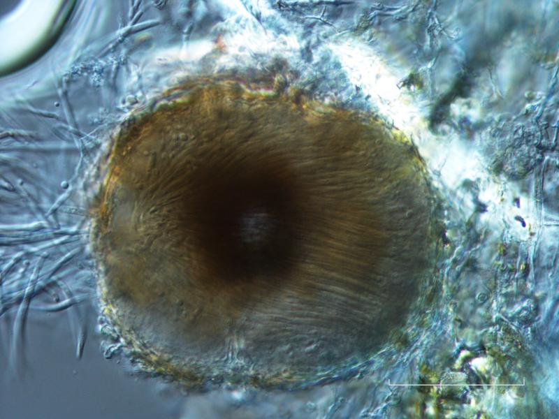
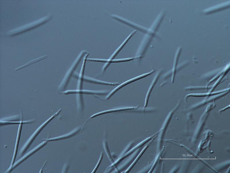
 Hello! I wanted to share a fascinating thing I saw this week. This is the coelomycete anamorph state of pyrenolichen Phyllocharis orbicularis (=Strigula orbicularis), which is apparently not illustrated anywhere. I was lucky enough to be able to ask Robert Lücking for help, and he ID'd it right away, having seen it before; when I asked where I could find an illustration, he admitted that he didn't think one existed, only the ascomata and the macromorphology of the thallus. The thallus of this particular example is somewhat poorly lichenized, and looks more like the photobiont (Cephaleuros virescens) than the typical thallus, but the conidia are the really fun part anyway.
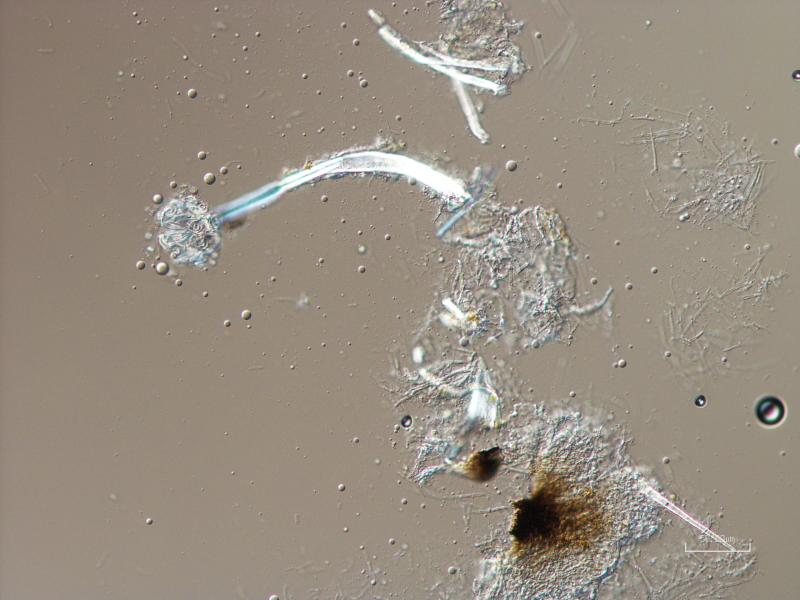
The conidia are hyaline, 4- to 6-septate, 40-45 x 2-3.5 ?m excluding the appendages, with a non-cellular, mucoid appendage at each end, which are quite variable in length, but generally less than 10 ?m, and often curving into a hook. Conidiophores are small, lageniform, reduced to conidiogenous cells, and integrated into the inner wall of the pycnidium.
I really wanted to put these photos out there, so that if anyone else is struggling to identify this beautiful and distinctive anamorph they'll be able to find some reference images! I can't thank Dr. Lücking enough for his kind help in the identification of this fungus.