12-03-2026 19:44
 Enrique Rubio
Hi to everybody.Can you give me any suggestions ab
05-03-2026 10:07
Hulda Caroline HolteHello, I found and collected this species growing
12-03-2026 15:45
Åge OterhalsDear forum,I found this small discomycete on a ver
11-03-2026 17:36
 Michel Hairaud
Bonjour, Je cherche des indices pour cette réc
14-03-2026 13:51
 Thierry Blondelle
Hi everybody Under Quercus ilex, i hesitate to na
08-03-2026 14:05
Thierry Blondelle
Bonjour à tous,Sur 3 récoltes supposées de H. l
11-03-2026 16:48
 Bruno Coué
Bonjour, je serais heureux d'avoir votre avis sur
Possible Orbilia cejpii
B Shelbourne,
31-03-2024 16:32
 • Orbilia, section Orbilia, key A: Asci inamyloid, yellow-orange apothecia, one globose SB with invisible filum, dead asci with strongly truncate apex in front view, hemispherical in profile view, narrow anchoring hyphae, low OCI, no gelatinous matrix, apical cells of paraphyses longer, tiny ellipsoid-(sub)globose spores, epithecium thin, sparse and granular, asci 8-spored, paraphyses uninflated to clavate-capitate.
• 'cejpii-eucalypti-tremulae' group: Deciduous wood, no glassy processes, carotenoid LBs in the paraphyses and excipulum, small ellipsoid-(sub)globose spores, small SBs, paraphyses strongly inflated and often asymmetric.
• Difficult to distinguish, possibly O. cejpii, or O. tremulae with short spores.
• Compared to O. eucalypti seen: Apothecia noticeably more yellow, excipulum yellow, exudate more yellow, spores smaller and more (sub)globose, anchoring hyphae more noticeable in macro.
• Temporary blue-grey reaction of material in IKI was very distinctive.
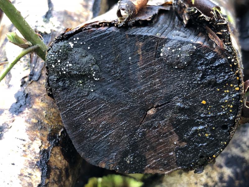
Habitat: On stromatic pyrenomycete and wood, on sawn end and decorticated parts of a medium-size log, looks like Betula pendula, in a pile of logs, within 0.5 m of the ground, damp at the time and possibly hygric, in a clearing, no bodies of water nearby, mixed deciduous woodland, mostly Quercus robur, southern England, late March.
Associates: Stromatic pyrenomycete, algae, anamorph with long brown multiseptate monoblastic conidiophores (presumably not from Orbiliaceae), Polydesmia pruinosa (on perithecia), Eriopezia caesia on a different log, several types of conidia found in mount.
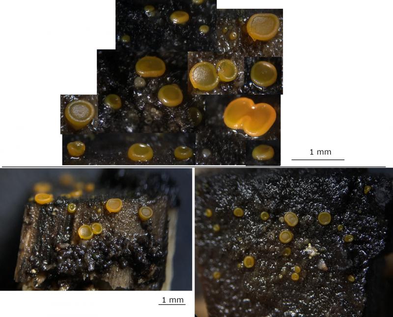
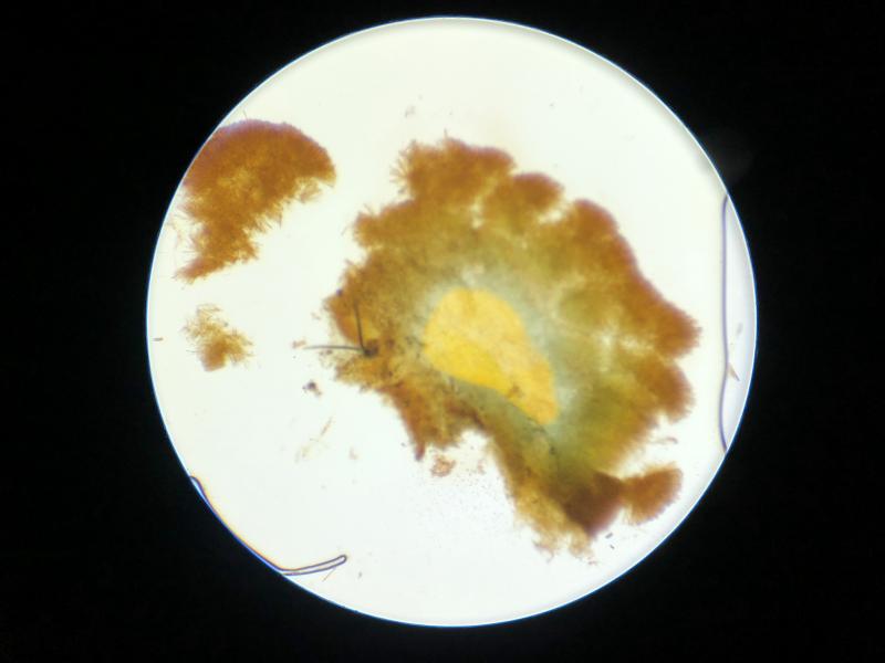
Apothecia: Group of ~30, gregarious to 2-caespitose, ~0.3-0.6 (1) mm diameter when mature, shallow-cupulate to discoid, mostly sessile but occasionally short stipe giving turbinate shape (Q < 1), superficial, appressed when sessile, pale yellow-orange to rose-yellow-orange (more light), +/- translucent, gelatinous appearance, texture firm-gelatinous, initially pulvinate-globose whitish to yellowish, receptacle opening early and then shallow cupulate; margin raised, toroidal, sometimes undulate, occasionally strongly lobate; disc more translucent (dark substrate showing slightly), plano-concave; anchoring hyphae around receptacle and base, more noticeable with stipe.
Storage and methods: Stored in a damp box for 48 hours, a median and an edge section taken from a single mature-looking apothecium, mounted in water, IKI added to water mount.
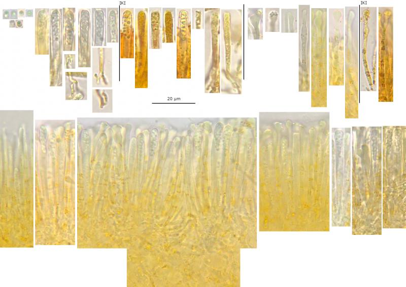
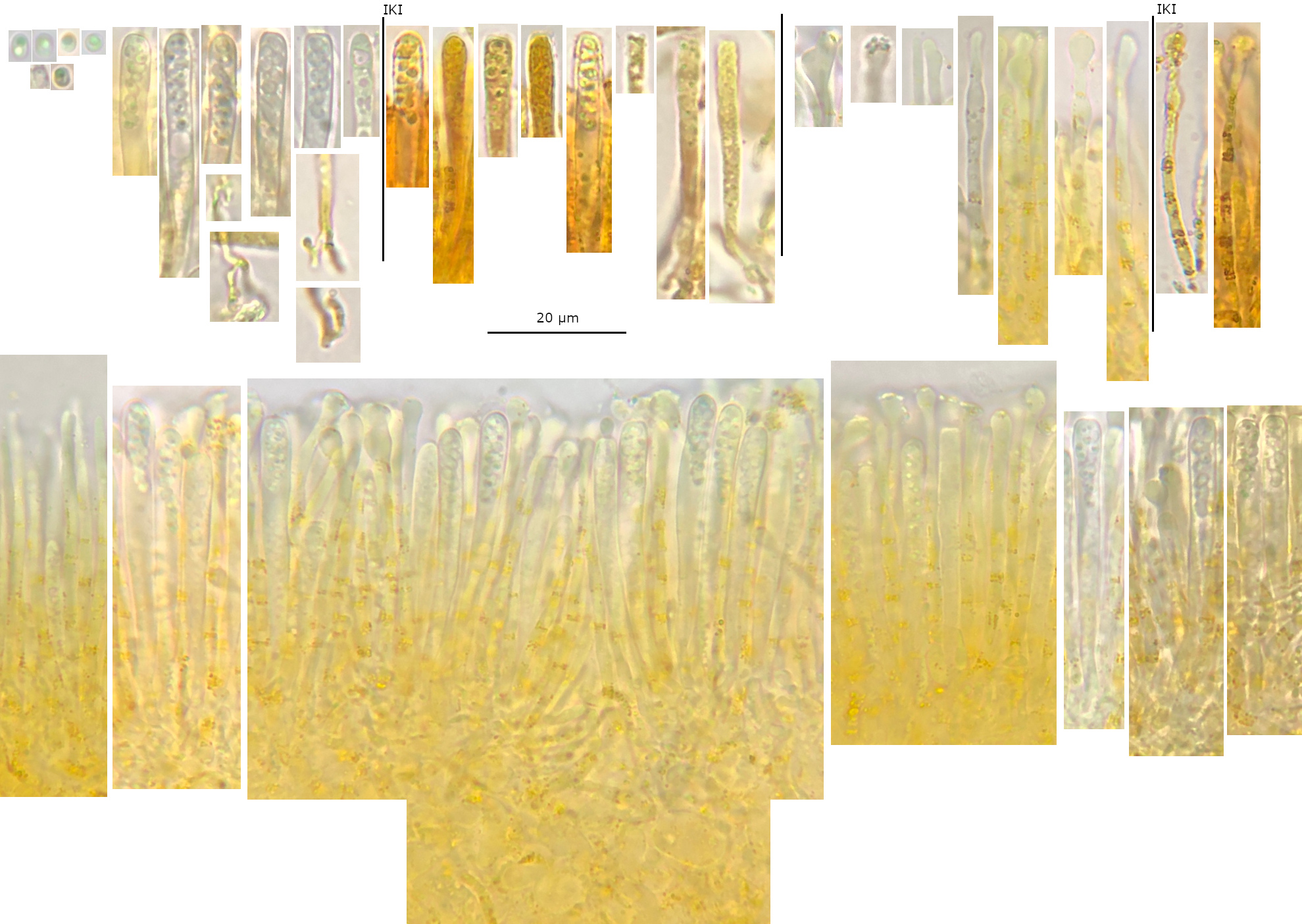
Asci: Cylindrical to slightly clavate, simple septa, bifurcate with H, h, y shapes observed, 1-1.5-seriate, spores variably oriented and more bunched at the apex, apparently very few discharging in water mount.
• Vital: 40-50 x (3.5) 4-4.5 µm, n = 9, apex rounded-obtuse, pars sporifera 10-20%, protruding above epithecium.
• Dead: (33) 35-45 (48) x 2.5-3.5 (4) µm, n = 10, limited loss of turgidity, eventually uniseriate, spores more loosely distributed throughout, apex truncate and often concave to forked in front view, hemispherical in side-view, small lens-shaped apical thickening.
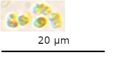
Spores: Measurements for vital and mature spores in water mount or asci – (2) 2.3-3.3 (3.5) x 1.4-2 (2.2) µm, n = 15, mean = 2.7 x 1.7 µm, Q = (1.3) 1.5-1.8, mean Q = 1.6, broadly-ellipsoid to subglobose, usually homopolar with ends rounded to obtuse, small to medium-size globose SB.
Paraphyses: 2-3 septate, sometimes branching at basal septa, apical cell usually longer 1.5-2x, near margin apex uninflated to slightly tapering ~1-1.5 µm wide, otherwise apically inflated ~2.5-4 (5) µm wide, clavate-capitate to irregular (e.g. additional subapical inflation, mammiform, asymmetric); LBs around septa and sometimes apex, carotenoid; VBs pale yellowish, large, slightly refractive.
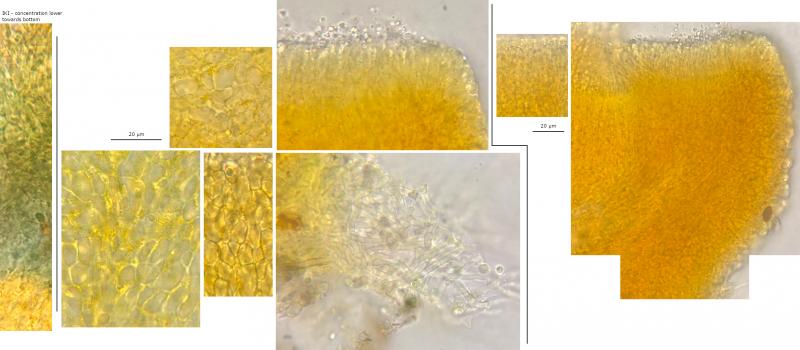
Exudate: Bright yellow to orange in concentration, clumpy around margin, thin and patchy epithecium, coating margin and flanks, grainy in surface view.
Subhymenium: Yellow with large swollen cells, carotenoid LBs.
Ectal excipulum: Yellow (at least at the surface), textura prismatica-globosa, slightly larger cells towards base, areas more strongly gelatinised, carotenoid LBs, rarely globose SCB in a cell, generally perpendicular to the surface, marginal cells smaller, broadly clavate.
Medullary: Yellow, divided by 3-4 horizontal hyphae, textura intracata-prismatica, seems weakly developed.
Basal attachment: Abundant anchoring hyphae around base and lower flanks, hyaline.

 Hymenium-0007.jpeg
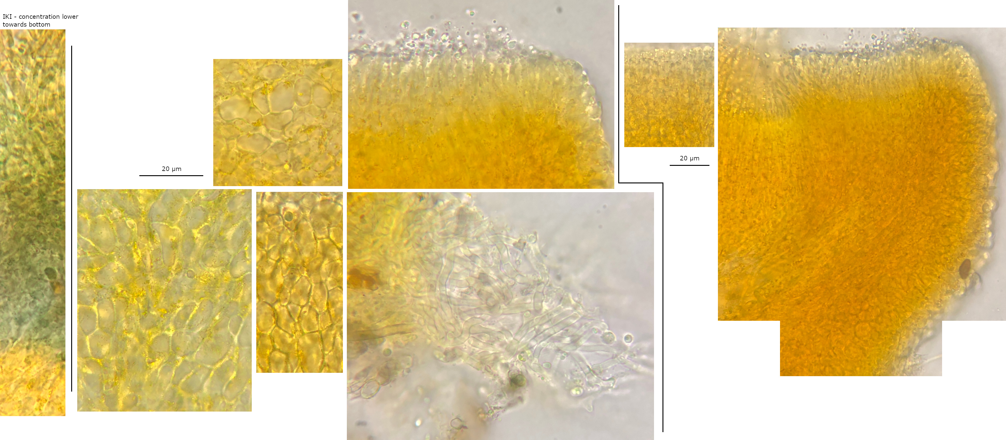 Excipulum-0002.jpeg
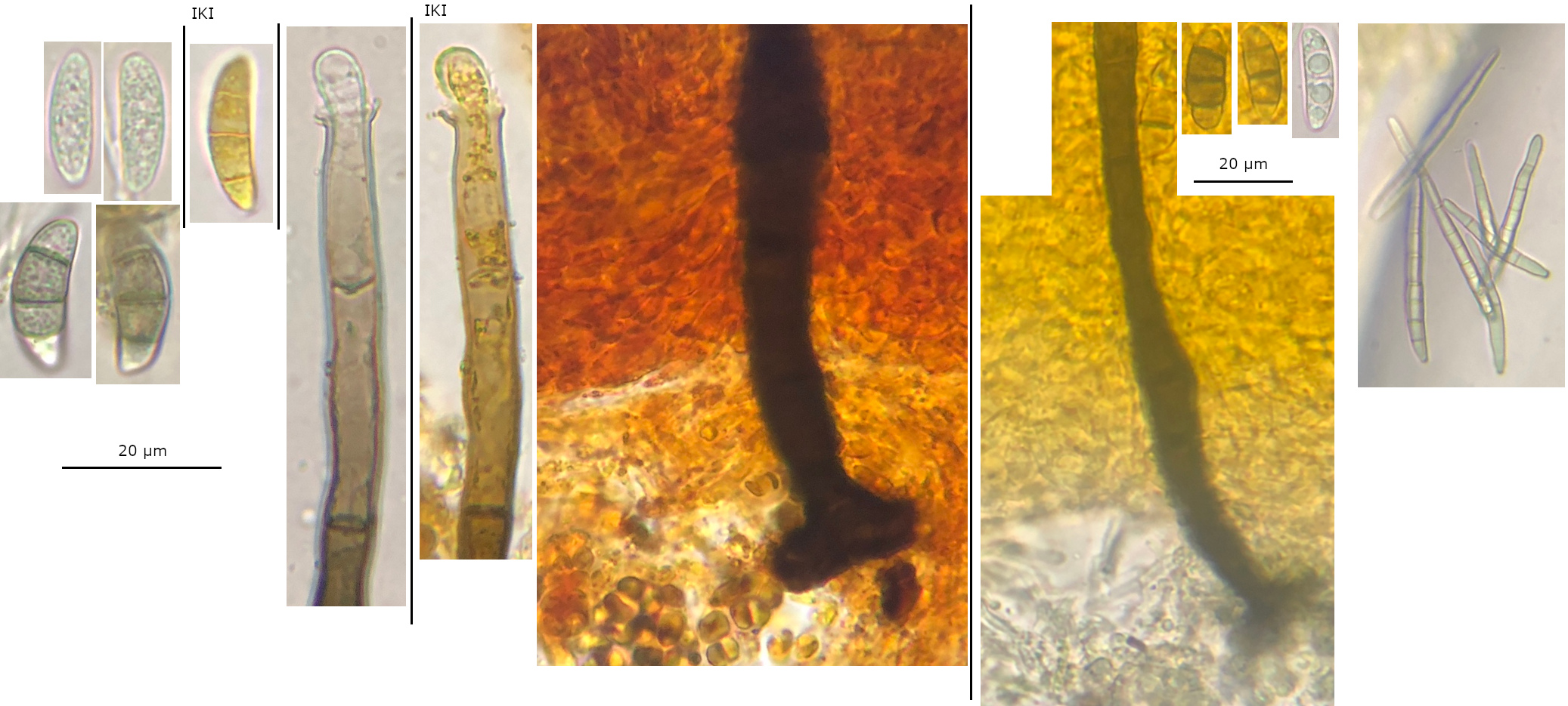 Anamorphs-unrelated-0001.jpeg
Hans-Otto Baral,
31-03-2024 21:13

Re : Possible Orbilia cejpii
This needs a higher resolution. Sounds interesting! The hypho is not unknown to me, but of course not related.
B Shelbourne,
31-03-2024 22:08
Re : Possible Orbilia cejpii
Sorry about the resolution, I have uploaded the original files now too.
I also include a 10x objective photo of the IKI reaction that I forgot.
I also include a 10x objective photo of the IKI reaction that I forgot.
Hans-Otto Baral,
31-03-2024 23:00
Re : Possible Orbilia cejpii
Now I understand. The free spores are doubtful. The spores you have inside the asci are clearly curved and warted: O. xanthostigma. The iodine reaction is due to the carotinoid.
B Shelbourne,
01-04-2024 00:18
Re : Possible Orbilia cejpii
Thank you, that explains a lot.
I should have looked at other groups in the section to understand how the spores differ, because now it is easy to see. The IKI reaction is also described for the xanthostigma-leucostigma complex.
I did previously notice that some spores looked a strange shape but with the overlapping then I thought that I was misintepreting it, and I must have thought the warts were small LBs. I think you're right about the previous free spores and I have included now some spores in IKI that were presumably discharged from an ascus, I was previously concerned that these were misshapen.