
11-06-2025 16:26
 Jason Karakehian
Hi everyone, I am looking for the following protol
17-06-2025 12:10
 Castillo Joseba
Del pasado dia 4 en bosque de hayas, en tierraA
12-03-2025 17:54
Karl Soler KinnerbäckHi all!Do you agree with Scutellinia hyperborea? P
17-06-2025 12:02
Castillo Joseba
Del pasado sabado, en bosque de hayas y avellanos,
13-06-2025 09:41
 Josep Torres
Hello.A cerebriform ascomycete sprouting scattered


 • Mollisia on tree leaves: On dead Quercus leaves, VBs in paraphyses (and elsewhere), and distinctive excipulum.
• Ingo's key (on asco-sonneburg) does not seem to include this substrate yet (point 1000), but in Zotto's folder, M. nervicola or M. aff. protrusa seem to be on Quercus.
• Itagaki et al (2019) describe collections assigned to 'Pyrenopeziza' nervicola and 'P.' protrusa from Japan, reporting the later to have smaller spores, longer and narrower asci, and less pigmented marginal cells.
• These authors reported sequences from these collections as distinct and uploaded sequences to GB, seem quite distant to any other Mollisia in a direct blast.
• Seems to be M. nervicola: Habitat, macro, spores, marginal cells.
• The paraphyses observed were mildly lanceolate when vital and fully turgid, and I noticed Mollisiopsis quercina that could be another synonym.
Habitat: In vitro on two dead leaves of Quercus robur, found after about three weeks in a damp box, leaves collected from leaf litter at the side of a path, mixed deciduous woodland, Low Weald, England, mid-April.
Associates: Brunnipila brunneola still fruiting on the leaves, possible pyrenomycete, small translucent fungicolous mites that appear to have flourished in the damp box. No clear sign that the mites have been eating the apos of the Mollisia sp., but certainly those of Brunnipila bruneola.
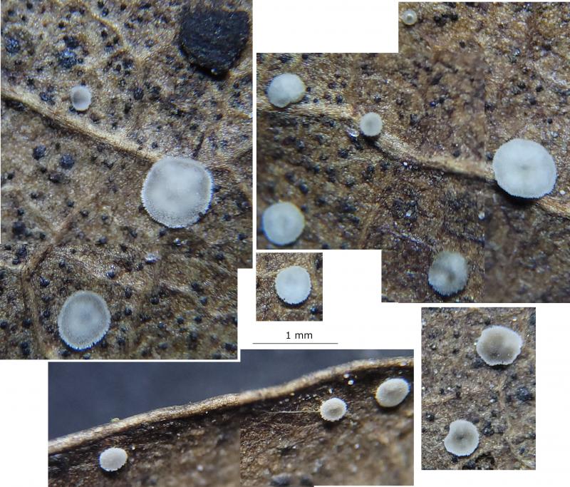
Apothecia: Many on each leaf, hypophyllous (on the underside), in gregarious groups, growing on the veins (and midrib), 0.3-0.5-0.9 mm diameter, superficial, initially whitish-greyish and cupulate, becoming discoid to pulvinate, appressed, with greyish-black patches – often a ring inside the margin or patch around the centre, soft-gelatinous texture, loosely attached to the substrate, possibly going black when over mature; receptacle initially greyish with a pruinose appearance (flank hairs?), darkening with age but only seen through the disc; margin with very short whitish hairs, initially raised but in maturity usually flat, round and mostly undulating slightly; disc whitish, translucent, pruinose/frosted appearance (protruding paraphyses), dark receptacle showing through in patches in maturity; no subiculum visible.
Storage and methods: A central section taken from a large and mature-looking apothecium, one day after discovery in-vitro, mounted in water, some pressure applied to separate the hymenium, IKI, KOH, and CR added in succession. A technical issue resulted in some problems with many photos taken, including those of more vital cells.
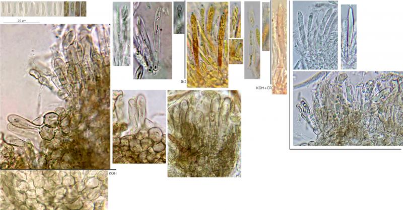
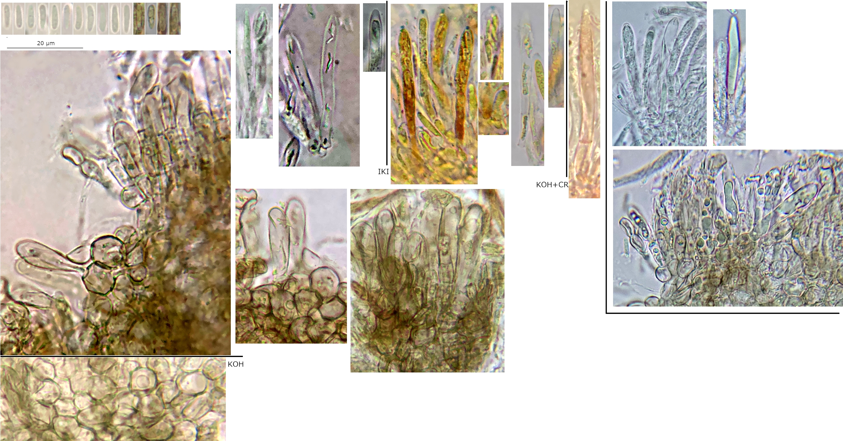
Asci: Cylindrical-clavate, apex round-acute, apical rings bb, croziers, 1-2-seriate.
• Vital mature: ~40-50 x 5-6 µm, pars sporifera ~30%, 2-seriate.
• Dead: ~30-40 x (2.5) 3-5 µm, pars sporifera >~70%, more cylindrical, apex more acute, usually with apical thickening ~1.5 µm.
Spores: Narrow ellipsoid-fusiform, rarely appearing allantoid, apices rounded-acute, usually heteropolar with the apex more rounded and the base more attenuated and acute, occasionally inequilateral – possibly in profile view, usually very slightly curved, apparently aseptate, usually 1-3 small shadowy LBs visible towards the poles or centre, LBs more noticeable when spores inside mature asci, often 2 whitish nuclei visible.
Vital spores measured in water or IKI, some in dead mature asci: (6) 6.2-7.2 (7.4) x 1.5-2.1 µm, Q = (3.2) 3.4-4.3 (5.4), n = 14, mean = 6.9 x 1.8 µm, Q mean = 3.9.
VBs: Yellowish-greenish, a single one filling apical cell of paraphyses, marginal cells, and cells of the ectal excipulum, very sensitive and quickly deforming when traumatised.
Paraphyses: ~40-50 x 3-4.5 µm, cylindrical-lanceolate, apex rounded-acute, 1-2-septate, apical cell ~2.5x longer, protruding above immature asci ~5-10 µm.
Exudate: Some largish, hyaline crystals, origin not clear.
Marginal cells: At the margin ~25-35 x 4-6 µm, at the flank ~15-22 x 6-7 µm, elongated, clavate, protruding part 0-2-septate, apical cell ~3-4x longer.
Ectal ex: Brownish, textura globosa, becoming slightly paler and greenish in KOH.
Subhymenium and medullary ex: Apparently hyaline, undeveloped, difficult to distinguish from each other.
Basal attachment: Not observed.
 Micro-2-0001.jpeg