12-06-2026 14:50
 François Freléchoux
Bonjour, Voici la brève description d'une Mollis
10-06-2026 21:16
François Freléchoux
Bonsoir,Le dernier du jour, en attendant votre avi
11-06-2026 19:01
 William Slosse
Hello all,In an attempt to make a culture of a sus
11-06-2026 19:03
 Nicolas VAN VOOREN
Chers membres d'Ascofrance,Le site sera placé en

09-06-2026 18:32
Camille MertensSur morceau de roseau immergé 0,5 - 0,7 mm de dia
10-06-2026 12:54
Steve ClementsBonjour encore, Pouvez-vous m'aider, s'il vous pl
10-06-2026 21:07
François Freléchoux
Toutes les tiges de gentianes jaunes de l'an pass�
10-06-2026 13:41
François Freléchoux
Bonjour à nouveau, Voici une trouvaille d'hier.
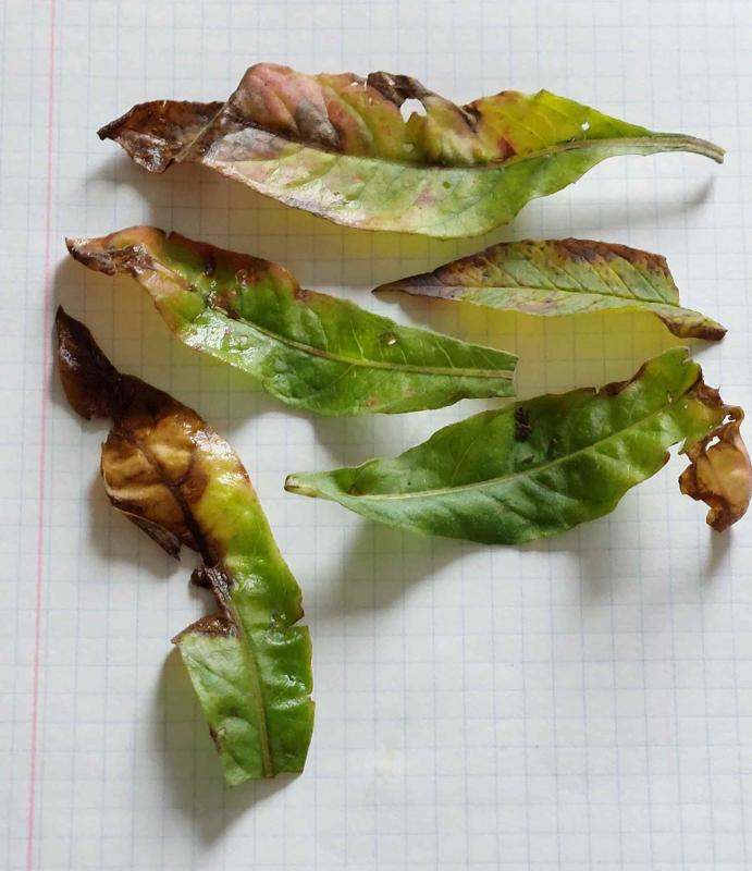
Conidiomata on leaves of Lysimachia vulgaris.
François Bartholomeeusen,
07-08-2024 18:24
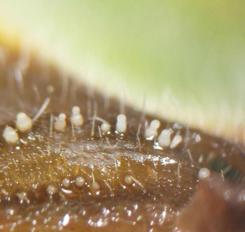
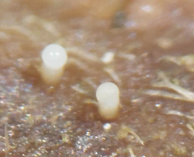
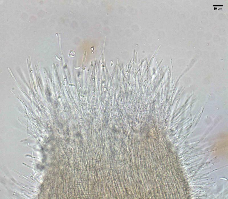
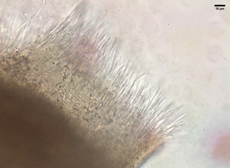
On the discoloured leaf lesions of Lysimachia vulgaris, caused by Septoria lysimachiae, I found several separately growing synnemata (white stem with a round droplet with secreted conidia at the top).
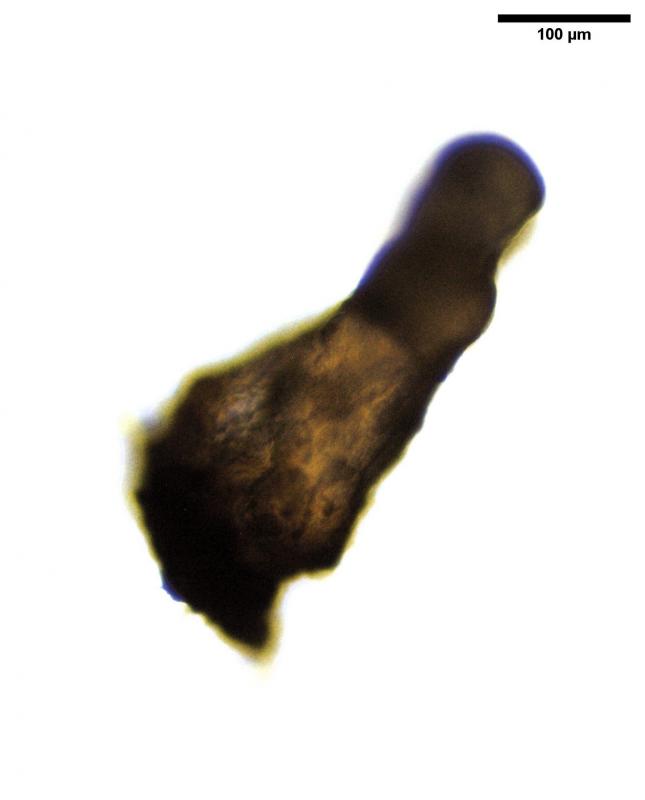
The fascicle has the following dimensions: height 410 µm, width base 175 µm, width top 93 µm
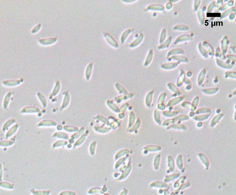
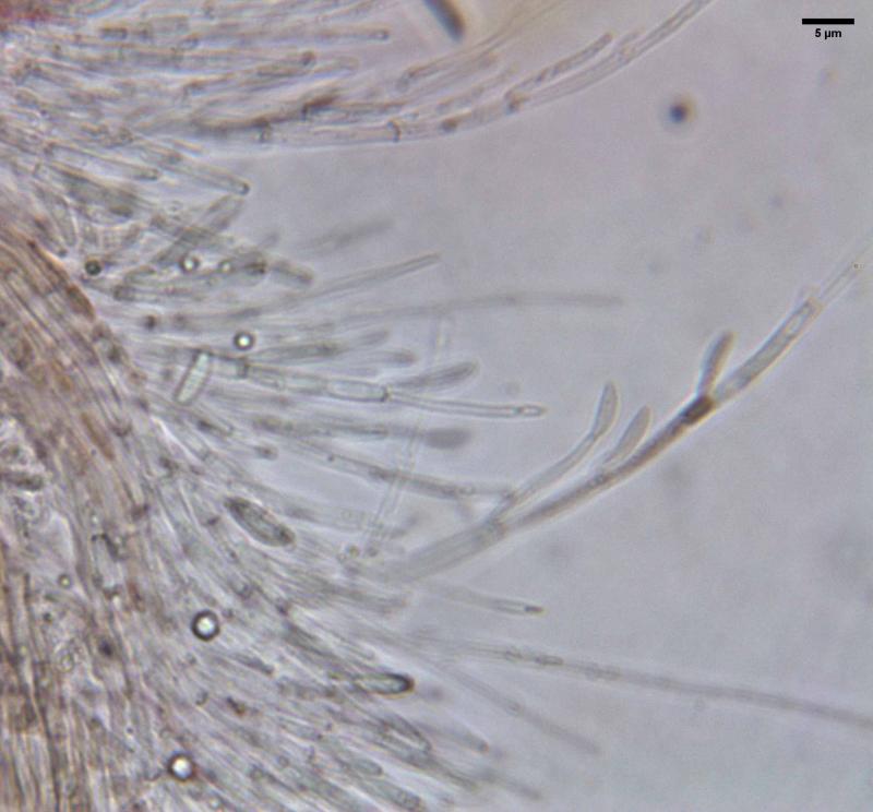
The conidiogenous cells are filamentous and have several branches. On them develop conidia, fusiform and in lateral view slightly allantoid: dimensions:
(5.2) 5.8 - 6.6 (6.9) × (1.5) 1.9 - 2.3 (2.4) µm; Me = 6.2 × 2.1 µm ; Qe = 3
Is there any connection with Septoria lysimachiae or is this an anamorphic form of another species. Any help is welcome as I am not coming to an identification.
Kind regards,
François