16-02-2026 21:25
 Andreas Millinger
Good evening,failed to find an idea for this fungu

08-12-2025 17:37
 Lothar Krieglsteiner
20.6.25, on branch of Abies infected and thickened

17-02-2026 17:26
 Nicolas Suberbielle
Bonjour à tous, Je recherche cette publication :

15-02-2026 04:32
One more specimen that is giving me some descent a

17-02-2026 13:41
Isabelle CharissouBonjour, est-ce que quelqu'un pourrait me fournir

16-02-2026 18:34
 Thierry Blondelle
Bonjour,La micro de cet anamorphe de Hercospora su

16-02-2026 17:14
Joanne TaylorLast week we published the following paper where w

16-02-2026 16:53
Isabelle CharissouBonjour, quelqu'un pourrait-il me transmettre un
• Mollisia suggested by excipulum and paraphyses.
• Of the species on wood and bark, M. ligni is reported on Tamarix and xeric habitats.
• M. ligni seems to fit, e.g. ascus rings, VBs, spores.
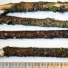
Habitat: On several low hanging branches of a Tamarix sp., still attached, growing from holes in the bark or decorticated parts, small and shrub-like plant, sheltered location behind sand dunes and close to a small building, West Beach, Angmering, south England, 26/11/24.
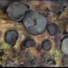
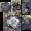
Apothecia: Gregarious to 6+-caespitose, blackish to dark grey to whitish, occasionally with a brownish tinge all over, diameter < 2 mm, initially globose-urceolate and blackish with a distinct whitish margin, then greyish, discoid and appressed, eventually more whitish and often convoluted.
Macro: Receptacle blackish with strongly pruinose or grainy appearance, sometimes more brownish at the flanks in maturity, margin distinctly whitish-translucent and remaining raised until maturity, disc initially dark grey and concave, becoming lighter and eventually whitish in patches, becoming plano-convex and eventually more convex and appressed, often developing deep wrinkles with the margin becoming strongly lobate and sometimes tearing, occasionally causing groups to appear cerebriform, apparently substipitate, sometimes associated with a black material on the substrate similar in appearance to the receptacle, especially in gaps in the bark, but this may be developing or failed apothecia.
Asci: Turgid ~55-65 x 5.8-6.5 um, croziers, rings IKI- but sometimes apex appears faintly bluish when turgid, 2-4-seriate.
Spores: Cylindrical-allantoid, slightly irregular, approximately homopolar but base often slightly more elongated, often with some tiny shadowy LBs towards each pole (not clear in photos), OCI 0-0.5.
Free living spores in water: (6.8) 7.6-9.4 (9.7) × (2.0) 2.1-2.4 µm, Q = (2.9) 3.4-4.2 (4.5), N = 30, mean = 8.5 × 2.2 µm, Q mean = 3.8.
Paraphyses: Cylindrical, apex not to gradually inflated, width ~2.75-3.5 (3.8) um, apical cell appears up to 3x longer, apex with several cylindrical to globose large VBs, yellowish en masse, apparently coalescing quickly and causing an empty space at the apex.
Medullary: Hyaline, text. porrecta, possible amyloid reaction noticed.
Ectal: Textura globosa, very dark at the surface, lighter below, brown hyphae seen in the substrate around the base.
Marginal cells: Cylindrical-clavate, brown at the flanks and hyaline at the margin, apex with a large cylindrical VB like the paraphyses, 0-2 septate.


The texture of the receptacle does seem distinctive. Personally, I prefer to the describe the appearance as densly granular rather than hairy, even if the protruding cells can be described as hairs.
I agree that the colour of the ascus apices is an artifact and I noted no reaction with the rings, probably due to my cheap objectives and phone camera. There is an interesting shape of the thickening, the oculus?, and the interaction with the top spore may encourage this artifact though. Upon review, I also think the greenish colour is not an amyloid reaction but some shadowing or overlapping with the dark patch seen in the attached photos.
I see that Le Gal and Mangenot (1956, 1958) describe a more or less extensive black stroma for the conidioma?, and a hypostroma for the apothecia. I also noticed some distinctive black material around the base of some apos, but this is not mentioned in your description, is this the anamorph?
Graddon's description and illustrations of Haglundia penyardensis seem to fit, it has inamyloid rings, and the measurements look good for dead cells. So, H. is a synonym of Mollisia (s.l.), because H. perelegans seems to be Mollisia (ligni var.) olivascens (and also Phialocephala urceolata). It looks like you also transferred H. elegantior to M. too.
In Tanney & Seifert (2020), the sequences available for M. ligni CBS 290.59 appear to be quite isolated in Mollisia (s.l.) and some of the morphology also seems quite isolated. In Ingo's key it is quite easy to identify apothecia as M. ligni, although this may not include some similar species.


 Hymenium-0033.jpeg