
12-06-2026 14:50
 François Freléchoux
Bonjour, Voici la brève description d'une Mollis
10-06-2026 21:16
François Freléchoux
Bonsoir,Le dernier du jour, en attendant votre avi
11-06-2026 19:01
 William Slosse
Hello all,In an attempt to make a culture of a sus
11-06-2026 19:03
 Nicolas VAN VOOREN
Chers membres d'Ascofrance,Le site sera placé en

09-06-2026 18:32
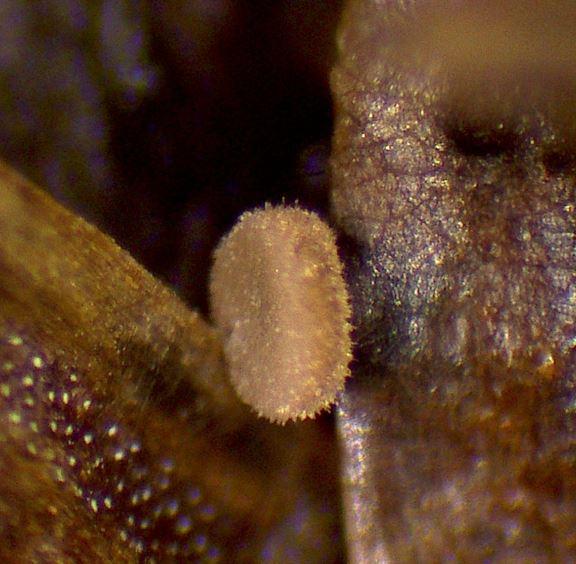
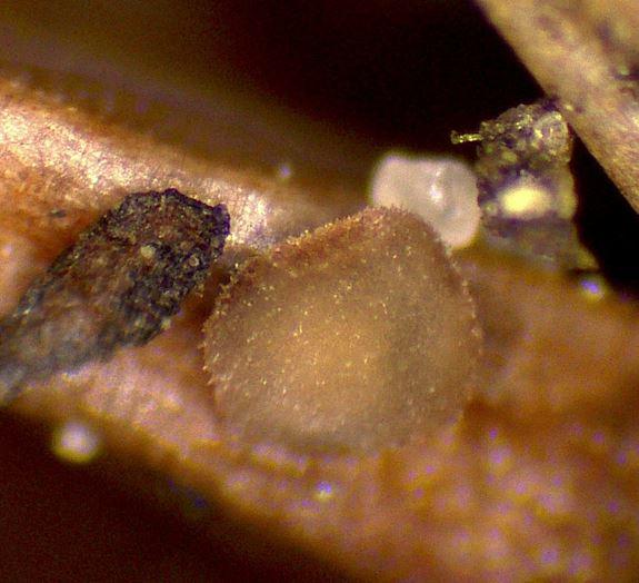
Camille MertensSur morceau de roseau immergé 0,5 - 0,7 mm de dia
10-06-2026 12:54
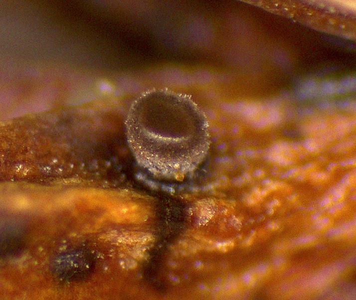
Steve ClementsBonjour encore, Pouvez-vous m'aider, s'il vous pl
10-06-2026 21:07
François Freléchoux
Toutes les tiges de gentianes jaunes de l'an pass�
10-06-2026 13:41
François Freléchoux
Bonjour à nouveau, Voici une trouvaille d'hier.
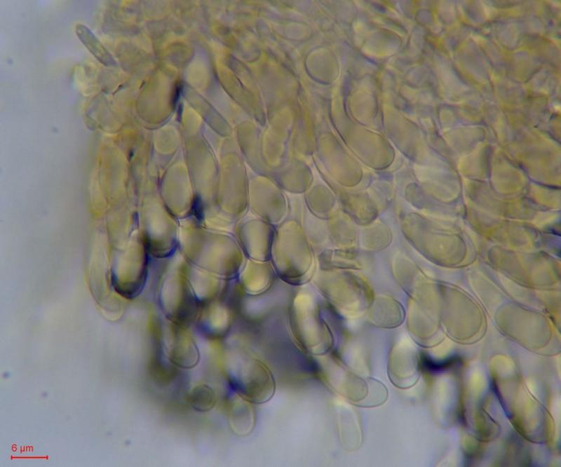
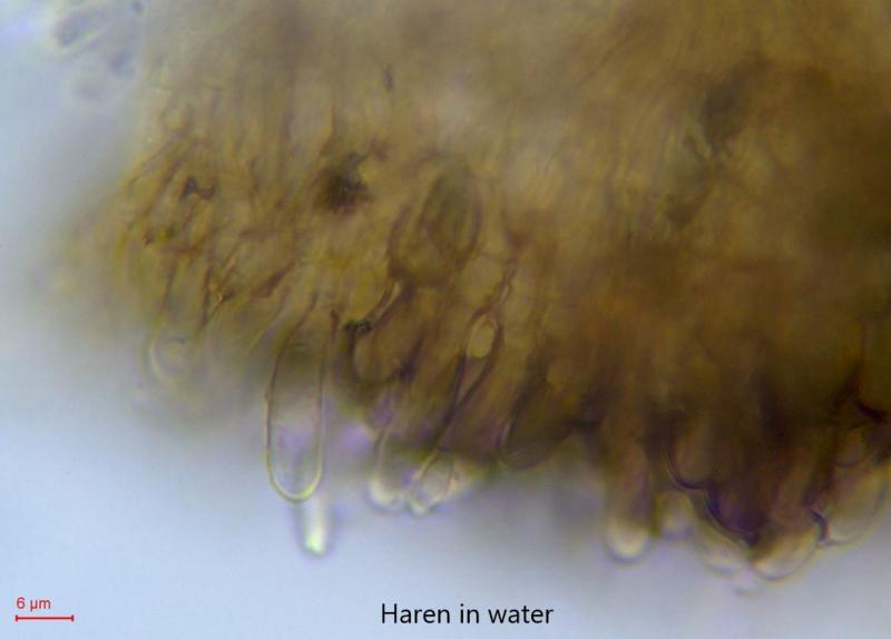
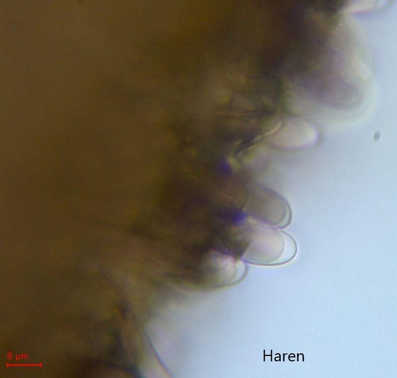
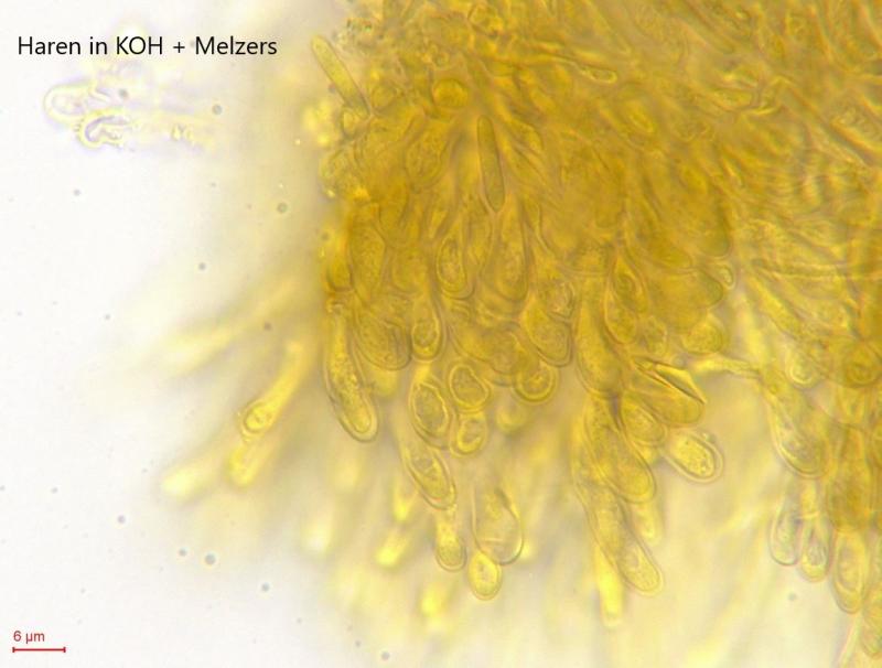
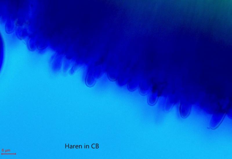
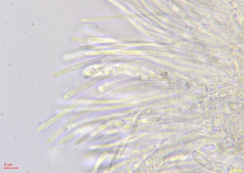
Hairs:
Smooth, no spines or warts, filled with VB/LB?
No change visible in KOH + Melzers or CB.
Spores:
Ellipsoid, somewhat more pointed towards the poles and small guttules at the poles, with 1 sept.
Measured in water: (9.8) 10.1 - 11.7 (12) × (3.1) 3.2 - 3.6 (3.9) µm; Qe = 3.3
Asci:
Measured in water: 64.2 - 72.4 × 5.5 - 7 µm; Qe = 11.5
IKI + and croziers +
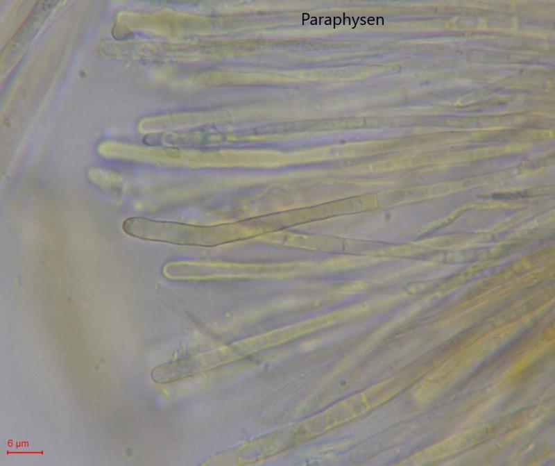
Paraphyses:
Cylindric, sometimes with a sept and somewhat constricted there, filled with VB's/LB?
Does anyone have an idea what this could be?
Many thanks in advance.
Kind regards,
Margot&Geert

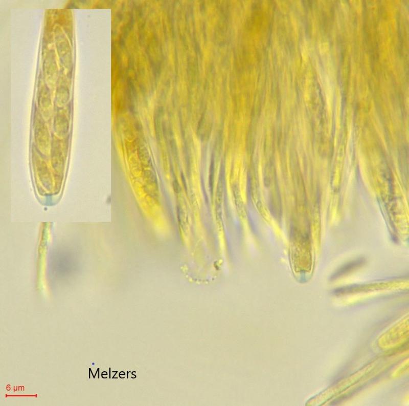

The photo with the J+ ascus is definitely in Melzers, if necessary we will repeat this in IKI.
Margot&Geert
We then looked at a younger disk in Melzers, I always make a preparation in water first, to see if it is good enough and then I indeed add a drop Melzers to the edge of the glass and suck it under, that is the photo you see here.

Melzer should better not used in discomycetes at all, for it is not necessary and causes cell death because of the chloral hydrate.
You should use IKI (lugol) or the higher concentrated "Barals solution" because it contains no toxic agents and lets the cells live.
Furthermore, hemiamloid reactions can only be examined in longer term with IKI, not with Melzers.
Yours, Lothar