
05-06-2026 11:02
Thomas Læssøehttps://svampe.databasen.org/observations/10596691
07-06-2026 15:10
 William Slosse
Hello everyone,On 05-06-26, I found following asco
07-06-2026 12:43
Steve ClementsBojour. This was a strange find on a stick on my
07-06-2026 12:09
 François Freléchoux
Bonjour, Voici une brève description de ce qui m
12-07-2015 00:05
 Nedim Jukic
This one from the same locality as the previous on
06-06-2026 17:44
Steve ClementsBonjour, This disco was on planed wood 3 x 1.5 cm
14-08-2016 23:15
 Alex Akulov
Dear friendsCan you help me to find the descriptio

04-06-2026 11:36
Gernot FriebesHi,found on Vaccinium myrtillus.Asci: IKI –, 8-s
05-06-2026 12:10
François Freléchoux
Capitotricha sp. sur Lonicea caerulea Caractères
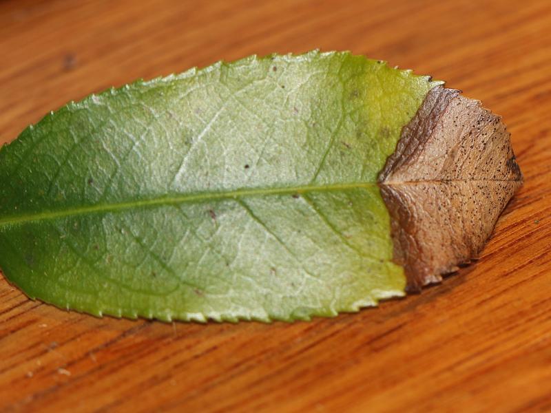
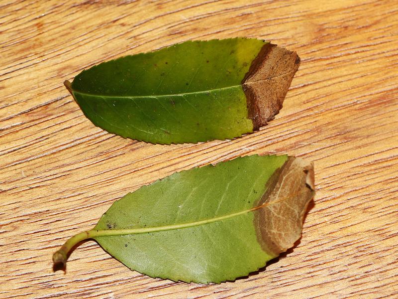
 On 6 february 2025 i stumbled upon a leafspot on Prunus lusitanica in Aerdenhout (The Netherlands). Can someone confirm that it is indeed Coleophoma prunicola? or maybe something completly different?
 For more photo's see: https://waarneming.nl/observation/338565267/
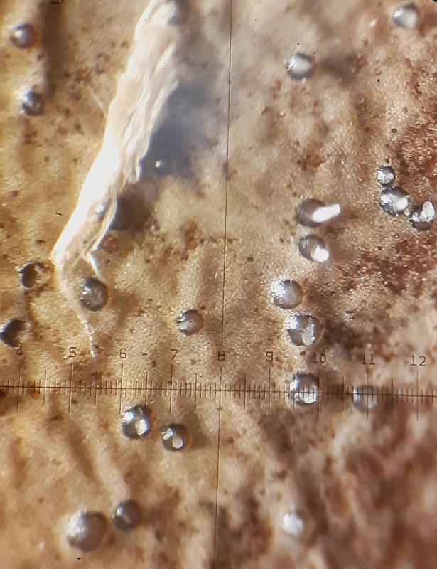
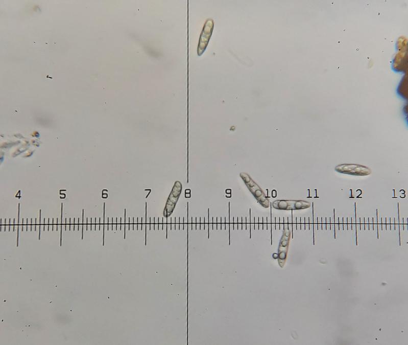
Conidiomate pycnidia, ca. 150- 200 µm diameter (N=10).
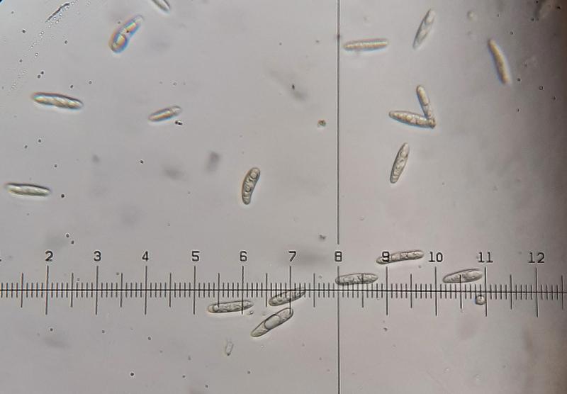
Conidia holoblastic, hyaline, aseptate, cylindrical, apex obtuse, base truncate, thin-walled, smooth, guttulate with several large guttules.
Conidia 20,8 - 23,4 - 26 ?m × 4-5,2 ?m (N=25)
400 X mafnification: 1 div. = 2,6 ?m
Following the Key for Coleophoma in the article: 'Reinstatement of Coleonaema for Coleophoma oleae and notes on Coleophoma' it should be Coleophoma prunicola
(Duan, J.X., Wu, W.P. and Liu, X.Z. (2007). Reinstatement of Coleonaema for Coleophoma oleae and notes on Coleophoma. Fungal Diversity 26: 187-204.) https://www.researchgate.net/publication/237440307_Reinstatement_of_Coleonaema_for_Coleophoma_oleae_and_notes_on_Coleophoma

To know this you should see the conidiogenous cells and compare them with those in the article.
Best wishes
Angel
Thanks for your comment. I can try another time to see the conidiogenous cells and compare them with those in the article. I already tried once but failed to see the conidiogenous cells sadly.
Kind regards,
Jorian