
29-04-2026 08:01
 Lothar Krieglsteiner
... on twig attached to small tree of Citrus auran
28-04-2026 22:51
 Bernard CLESSE
Bonsoir à toutes et tous,Pourriez-vous m'aider Ã
28-04-2026 20:33
Vitus SchÃĪfftleinHello, I found Trochila ilicina on Ilex aquifoliu

28-04-2026 21:50
 Pablo Sandoval
Hola a todos,Espero se encuentren bien. Hace mucho
27-04-2026 18:05
Lothar Krieglsteiner
... still attached at standing tree. The green con
28-04-2026 20:07
Lothar Krieglsteiner
... on twig in the air at standing Ceratonia siliq
27-04-2026 20:52
Lothar Krieglsteiner
Found on hanging tiwg of Olea europaea in dried-ou
27-04-2026 18:48
Tony MoverleyCollected 23rd April 2026, Norfolk, EnglandSwarms
27-04-2026 17:41
Lothar Krieglsteiner
.. Algarve, same leaf than the last post. The con
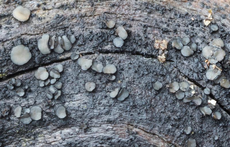
 Good afternoon
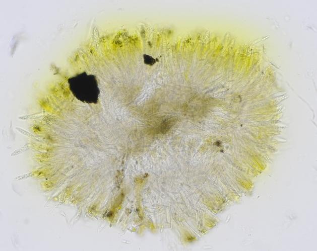
This 1-2 mm Mollisia was growing on Cistus ladanifer wood and I can't find any that look like it.
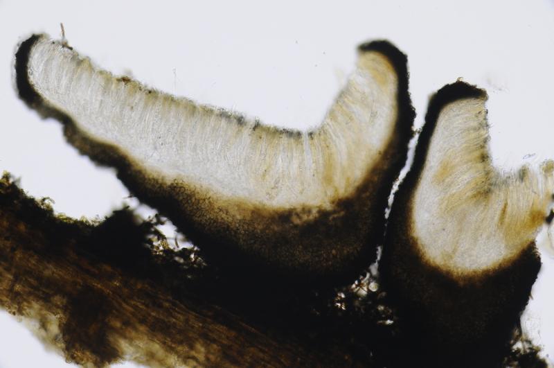
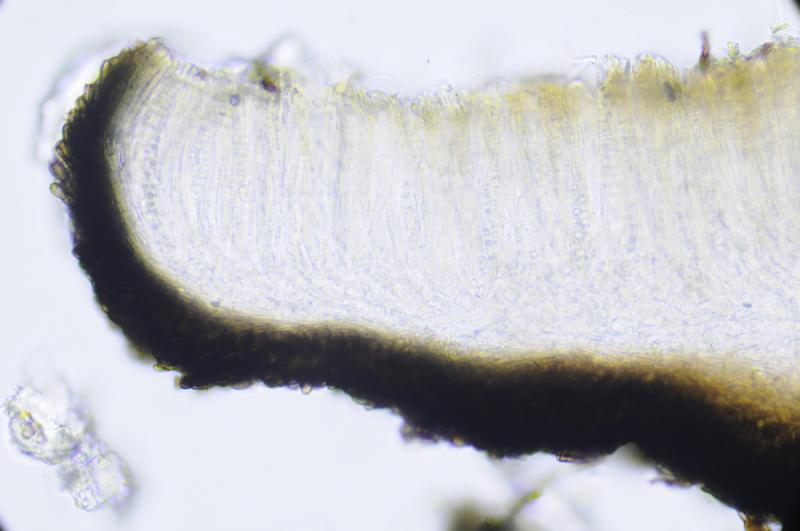
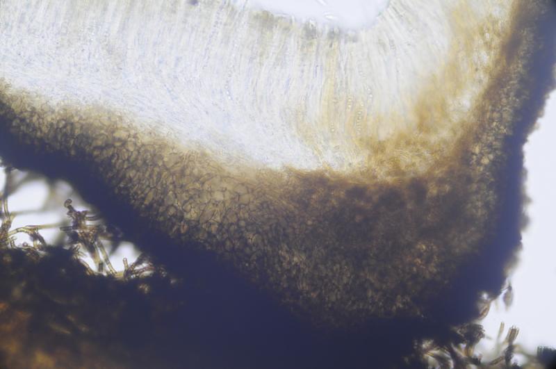
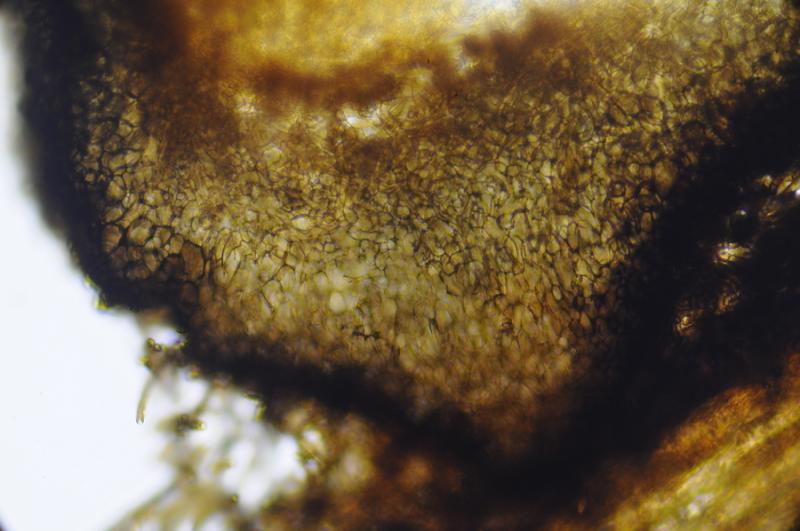
Ectal excipulum brown with textura prismatica to angularis, with claviform terminal cells. Medullary excipulum hyaline with textura prismatica to intricata.
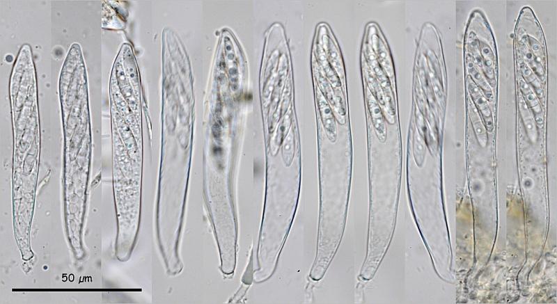
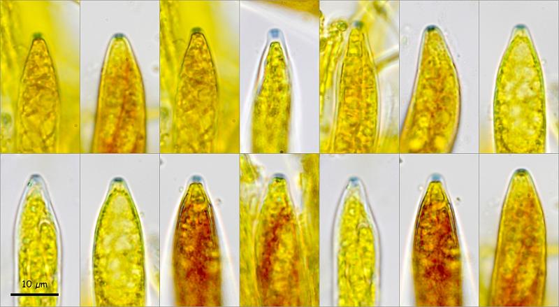
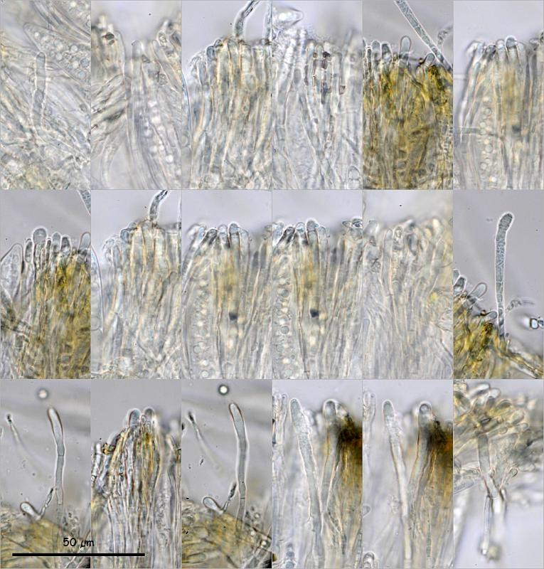
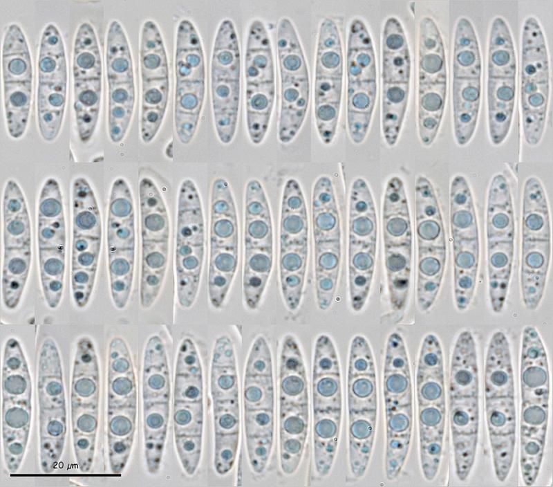
Asci octosporic, biseriate, with croziers, IKI+. Spores cylindrical-fusiform, usually asymmetric, with one side flatter and the other more convex, up to 3 septa, even inside the asci, with 1 large LB in each cell, at least in the 2 central cells. Paraphyses cylindrical, somewhat thickened at apex, with a large VB occupying the entire terminal cell, KOH+ intense yellow, with brown epithecium.
Asci: (76) 83.9 - 106.5 (111.9) Ã (10.3) 11.6 - 13.3 (13.9) Âĩm, Me = 95.5 Ã 12.3 Âĩm
Spores: (20.3) 21.8 - 24.9 (28.6) Ã (4.1) 4.6 - 5.2 (5.6) Âĩm, Q = (3.9) 4.4 - 5.3 (6) ; N = 66; Me = 23.6 Ã 4.9 Âĩm ; Qe = 4.8
I can find no Mollisia, Pyrenopeziza, Belonidium or similar with these spores. The closest spores I can find are those of M. ventosa. The spores of M. pilosa also have 3 septa, but they are larger and the asci are rr. I have also seen a picture of M. minutella with similar spores, but not the same.
Thanks for your help.
Miguel Ã. Ribes
Thanks

Very kind.Â
Best wishes.