
27-04-2026 20:52
 Lothar Krieglsteiner
Found on hanging tiwg of Olea europaea in dried-ou
27-04-2026 18:48
Tony MoverleyCollected 23rd April 2026, Norfolk, EnglandSwarms
27-04-2026 17:41
Lothar Krieglsteiner
.. Algarve, same leaf than the last post. The con
27-04-2026 18:05
Lothar Krieglsteiner
... still attached at standing tree. The green con
27-04-2026 17:16
Lothar Krieglsteiner
.. Algarve, moist lying.The conidiomata look like
27-04-2026 12:54
Steve ClementsBonjour. Ce petit champignon blanc résupiné et
27-04-2026 09:59
Pauline. PennaBonjour Can anyone advise me on these pycnidia fo

22-04-2026 20:54
 Enrique Rubio
Hi to everybody.This Pyrenopeziza grew in moist le
Discomycete on leaves
Josep Torres,
02-10-2025 09:16
 Hello.
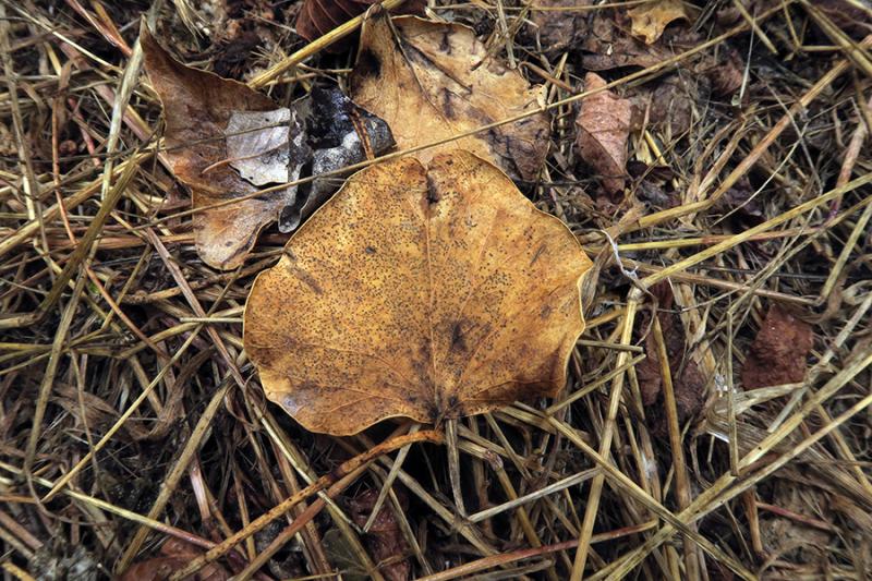
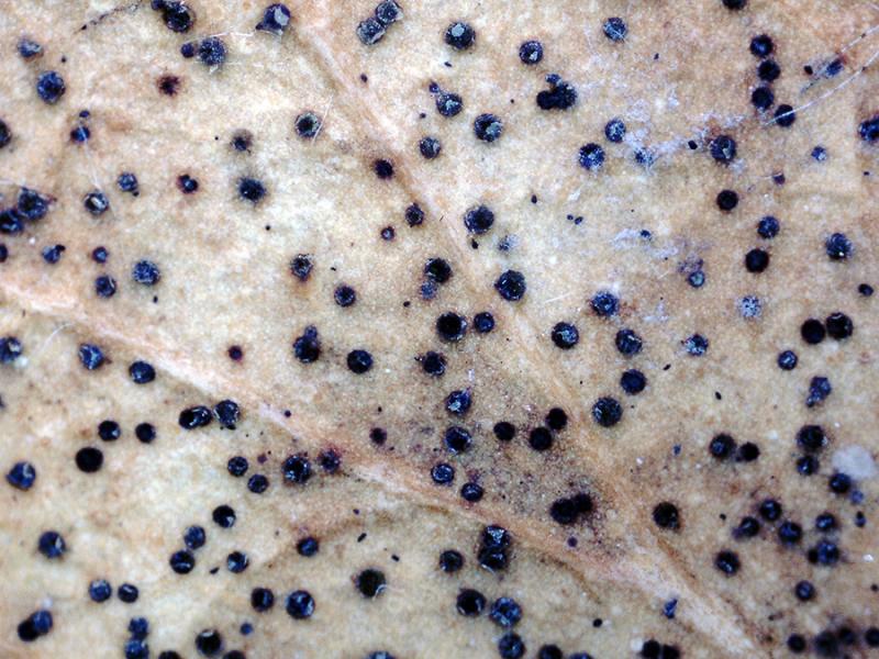
Some tiny apothecia sprouting in a scattered but abundant manner on decaying leaves in the riverine forest under poplars (Populus).
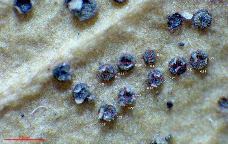
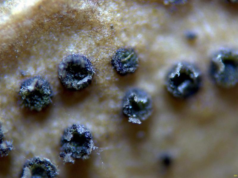
The apothecia are blackish, only 0.2 to 0.3 mm in diameter, without a clearly distinct margin.
Under microscopy, I couldn't see any structures that could correspond to marginal hyphae; if they were present, they would be very few.
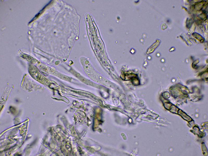
The excipule is very sparse, globose in texture, angular.
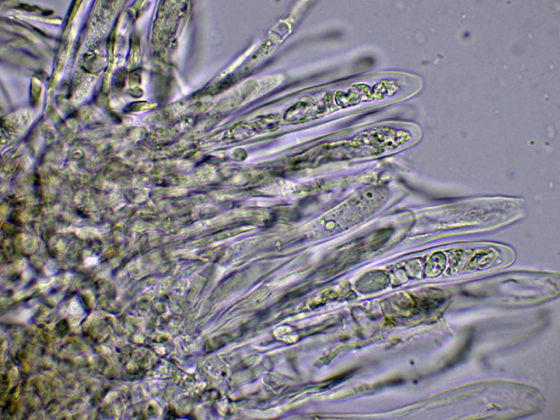
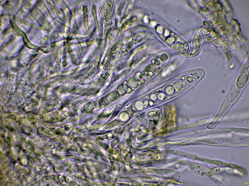
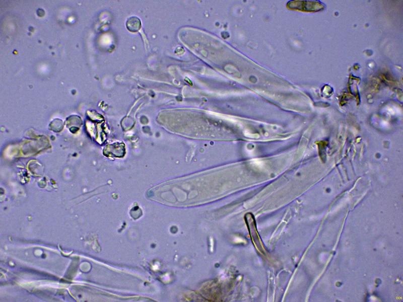
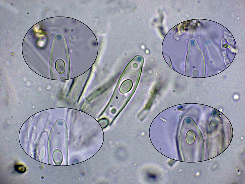
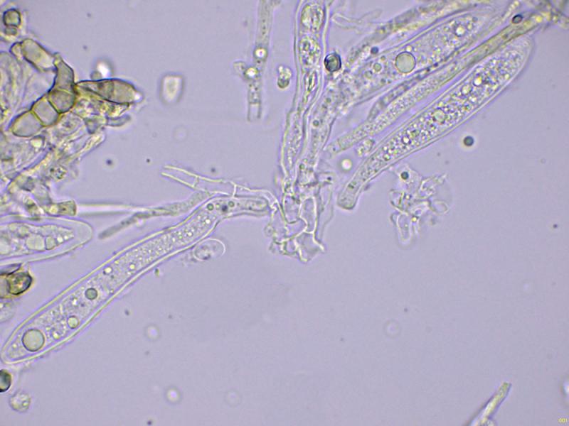
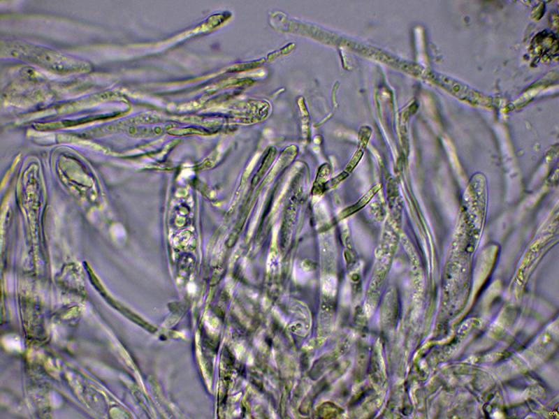
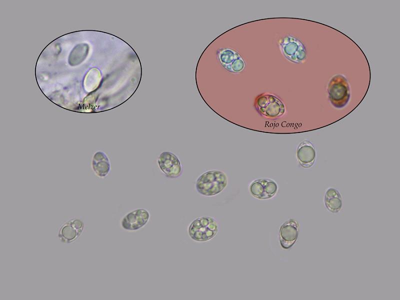
Octosporic asci, uniseriate, thick-walled, apparently without clear uncinules at their base, with measurements in water of (50.1) 56.1 - 70.8 (79.2) × (8.6) 9.3 - 11.4 (11.6) µm., Me = 64.9 × 10.4 µm., and with an amyloid apical apparatus to Melzer, cylindrical to slightly widened at its upper part, with measurements of 2.6 - 3.1 (3.4) × (2.1) 2.12 - 2.29 (2.3) µm. As a curiosity, I could not observe this apical apparatus in the preparations with Lugol. The paraphyses are filiform, septate, with a slight widening at the apex, protruding above the level of the asci.
Ellipsoidal ascospores, with one or more lipid droplets inside, inamyloid in the Melzer test, and with measurements in water of (8.2) 8.8 - 11.2 (11.4) × (5.4) 5.9 - 8 (8.4) µm, Me = 10 × 6.8 µm; Qe = 1.5.
Based on microscopy and the amyloid reaction of its apical apparatus, the closest I could find would be Drepanopeziza, but its appearance doesn't match.
Any feedback from you would be welcome.
Thank you in advance.
Best regards.
Josep Torres,
02-10-2025 09:18
Re : Discomycete on leaves
The rest of the images.
Hans-Otto Baral,
02-10-2025 09:23

Re : Discomycete on leaves
You have Trochila craterium on Hedera leaves I think, unless these are not Ilex leaves.
Josep Torres,
02-10-2025 09:26
Re : Discomycete on leaves
Thank you, Zotto, for your prompt response. Ivy (Hedera helix) is abundant in the area, and it's most likely the Trochila craterium you're suggesting.
Best regards.
Best regards.