
04-06-2026 10:50
 François Freléchoux
Bonjour, J'ai trouvé hier un petit asco observé
04-06-2026 07:02
François Freléchoux
Bonjour, Voici la description d'une espèce qui p
04-06-2026 13:34
Gernot FriebesHi,I am interested to hear your opinion on this Le
04-06-2026 11:36
Gernot FriebesHi,found on Vaccinium myrtillus.Asci: IKI –, 8-s
22-05-2026 13:29
Gernot FriebesHi,I am curious to hear your opinion on this mater
18-10-2022 00:12
Valencia Lopez Francisco JavierHola amigos/asRecientemente encontré esta colecci
03-06-2026 19:45
 Miguel Ángel Ribes
Good afternoonI'm completely baffled by this suppo
03-06-2026 14:39
Thomas FlammerApothecia yellow, glassy-transparent, 80 - 120 ymS
Hymenoscyphus?
Josep Torres,
23-12-2025 08:27
 Hello.
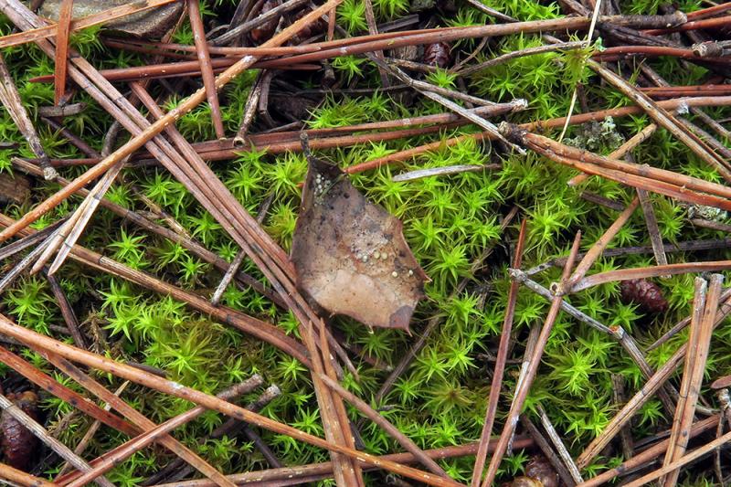
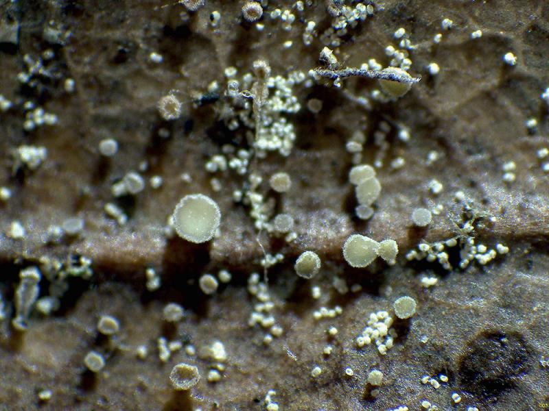
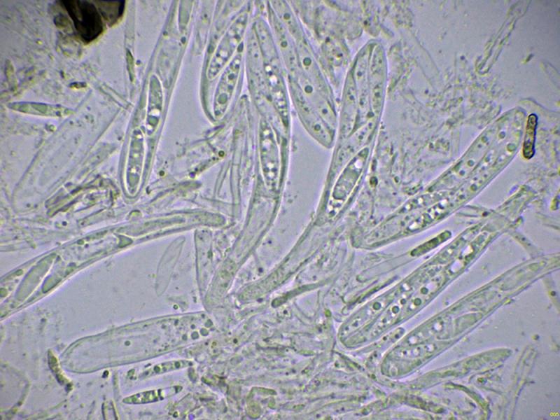
Small, yellowish ascomata, with very short and rudimentary stalks, greenish in mature specimens, sprouting from the surface of decaying leaves of Quercus rotundifolia.
Only 0.2 to 0.4 mm in diameter.
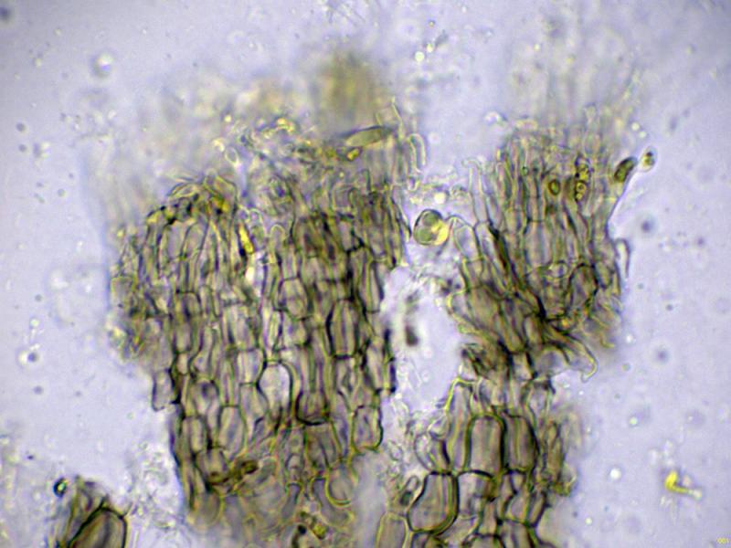
Basal hyphae elongated, arranged parallel to each other, and pigmented.
Hyphae of the excipulum globose-angularis in texture.
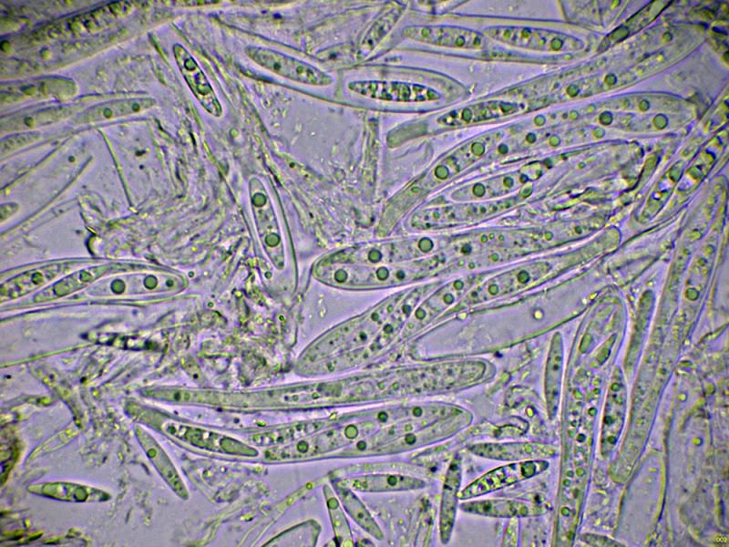
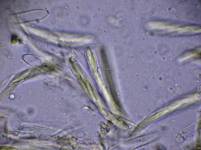
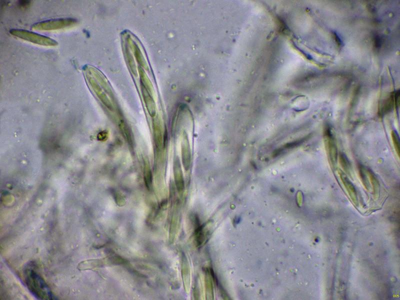
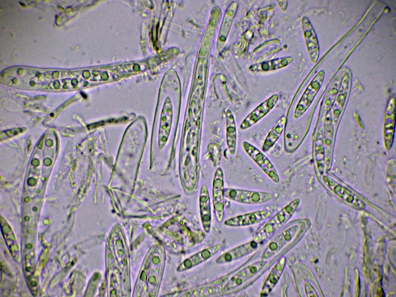
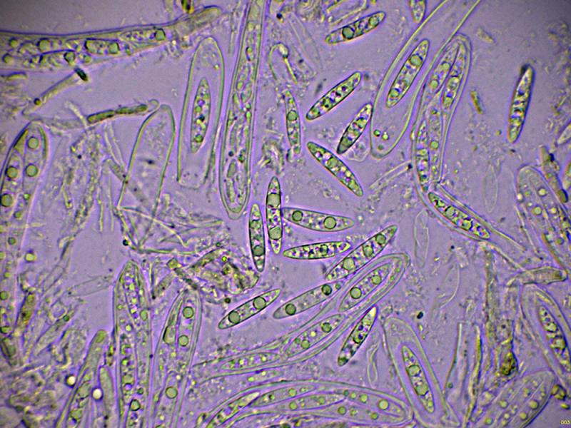
Octosporous asci, with an amyloid reaction in their apical apparatus, and although difficult to observe in some of these asci, I thought I saw croziers. Fusiform ascospores with pointed ends, two or three large lipid droplets and several smaller ones scattered throughout, measuring in water:
(14.3) 16 - 19.3 (22) × (2.8) 3.1 - 4.3 (4.5) µm
Q = (4.2) 4.3 - 5.5 (5.7) ; N = 40
Me = 17.8 × 3.6 µm ; Qe = 4.9
Based on these characteristics, I think it could be a species of the Hymenoscyphus/Phaeohelotium complex, but I have no other suggestion that convinces me.
Any opinions you may have would be welcome.
Thank you in advance.
Best regards.


Hans-Otto Baral,
23-12-2025 10:57

Re : Hymenoscyphus?
You don't have living paraphyses? That would be helpful. Surely no Hymenoscyphus, the apical ring is more of the Calycina type. The brown excipulum is remarkable. There are no hairs?
Josep Torres,
23-12-2025 14:22
Re : Hymenoscyphus?
Thanks, Zotto.
I couldn't observe any structures in the prepared samples that could correspond to hairs. The closest thing were the terminal hyphae visible above the image of the excipulum, which might correspond to marginal hyphae. There were no live paraphyses either. The apothecia were past their prime, and there were only asci and a collapse of spores. Since I have quite a bit of material from this sample, if we can't reach any conclusions, I'll send it for sequencing in my next shipment, which would be next year.
Best regards.
Hans-Otto Baral,
23-12-2025 14:57
Re : Hymenoscyphus?
That would be interesting to obtain DNA. It looks a bit like a Hyphodiscus. But such big spores are unknown there.
Josep Torres,
07-03-2026 22:46
Re : Hymenoscyphus?
Hi Zotto.
The sequence came back slightly noisy and didn't clarify much, but the ITS gene sequencing points with a 97.66% match to Calycellina sp., or at most Pezicula.
Best regards.
The sequence came back slightly noisy and didn't clarify much, but the ITS gene sequencing points with a 97.66% match to Calycellina sp., or at most Pezicula.
Best regards.
Hans-Otto Baral,
08-03-2026 08:34
Re : Hymenoscyphus?
Could you please send me the sequence and especially the chromatogram (ab1), then I will try my alignments wit partly unpublished sequences.
Josep Torres,
08-03-2026 14:35
Re : Hymenoscyphus?
Hi Zotto.
They're already in your email.
Best regards. Josep
They're already in your email.
Best regards. Josep
Hans-Otto Baral,
08-03-2026 21:29
Re : Hymenoscyphus?
Thanks, yes, the sequence is noisy, but only at a few positions. The result is clear: It must be a Pezizellaceae. Pezicula is a misID.
I tested ITS and S1506 intron separately:
ITS: 98.5% "Pezicula sp." UDB04294927, 98% Mollisina sp. UDB04221998, 97.8% UDB04222001, many Mollisina sp. 97.4% UDB, 96% Scleropezicula alnicola,
Intron: 91% Helotiales sp. LC064903, 86% Calycellina fagina, 81% Nagrajchalara
I think I see marginal hairs with short somewhat hooked whip. I am also sure there have been VBs inside the hair cells. So Mollisina or especially Calycellina are good options.