
20-05-2026 12:57
 Ingo Ibelshäuser
Hello everybody, on decayed hardwood e.g. Quercus
22-04-2026 20:54
 Enrique Rubio
Hi to everybody.This Pyrenopeziza grew in moist le


19-05-2026 12:55
 Hardware Tony
After checking Gminder and Otto's library I cannot
19-05-2026 10:27
Patrice TANCHAUDBonjour, récolte récente sur terre retournée i
18-05-2026 12:43
Sylvie Le GoffBonjour à tousPuis je avoir votre aide sur ce que
19-05-2026 14:56
Åge OterhalsI found this white cushion-formed ascomycete on ro

18-05-2026 10:13
 Lieve Deceuninck
Dear forum members,I identified this as the teleom
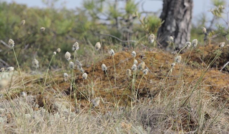
A search through cottongrass (Eriophorum vaginatum) litter yielded 8 morpho-species, half of them in anamorphic stage, and 5 are new in the bog list.
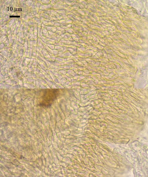
Small yellow discs from Hyaloscyphaceae were abundant at most leaves in wet locations. The hairs at outer surface and at the edge are very delicate, and soon disappearing on drying.
(#4176 - https://www.cubby.com/pl/%234176/_b9a126639359405abf5907978b8fac85)
Another is tiny perithecia of some Mycosphaerella forming in dense areas under leaf epidermis, look as black spots under the lens, and quite difficult to work with (inspite that i have the thinnest needle). But they are rewardetly fertile, whith short fissitunicate asci and hyaline two-celled spores inside.
(#4178 - https://www.cubby.com/pl/%234178/_86c50267b65a47f3a517f30c71e4cb15)
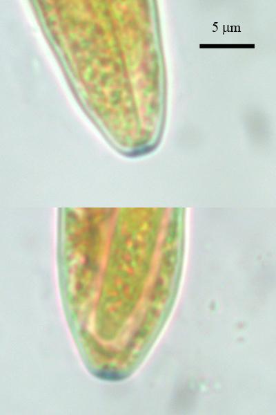
Third collection is interesting to see at Eriophorum, since i saw a similar species (Odontotrema) on bog pine wood. I suppose that it is some relative, e.g. a member of Ostropalean group since its characteristic asci with amyloid apical part.
(#4182 - https://www.cubby.com/pl/%234182/_bfa82e8746794d74aeeee43146bfaba0)
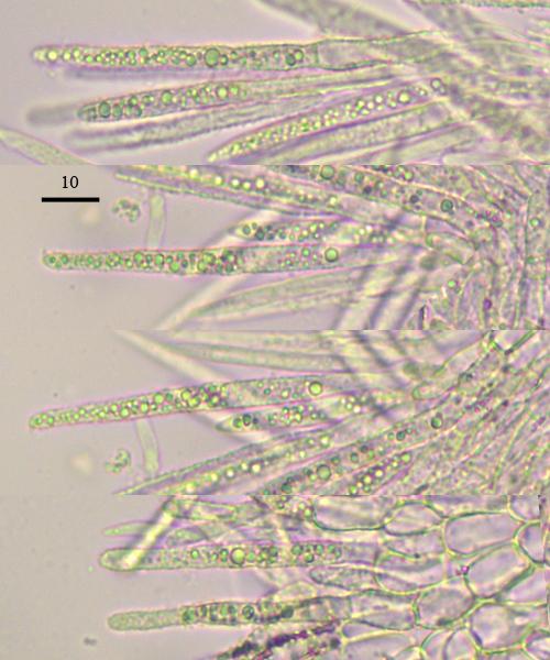
Lachnum sp. was met several times, it have long fusoid spores, probably i had identified this species already but it still need to be worked out with the literature.
(#4184 - https://www.cubby.com/pl/%234184/_cdf7fc8d2cb6474c8f64e4835e44f3f1)
And now the anamorphic species. Most commonly seen at old destroyed leaves are flat black conidiomas, where cylindrical conidia develop from inside ampulloid cells.
(#4177 - https://www.cubby.com/pl/%234177/_a14d3aabf4f743919fc40272c5d74d91)
Another one from Sphaeropsidaceae. Large septate conidia are formed from inside large globose cells placed inside of the walls of pycnidia.
(#4179 - https://www.cubby.com/pl/%234179/_a8a81e1cabdf453493bf273f8367a35e)
Demateaceous hyphomycete in form of brown bushes are developing at upper parts of the leaves.
(#4181 - https://www.cubby.com/pl/%234181/_638f66148728401b85c1490bd2998816)
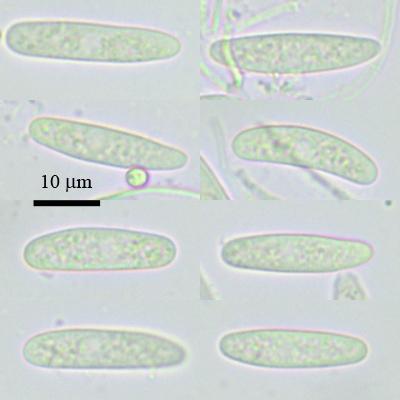
And the last conidioma were spotted accidentially with another species in the mount. It is someone sporodochia-forming with spherical phyaloconidia and ampulliform conigiogenous cells.
(#4180 - https://www.cubby.com/pl/%234180/_94a7c33c675d42929d94e97eb982ad4f)

Did you measure the spores? I would lke to see a photo of them, also of the hairs, but I have no time to scroll to your many images.
Zotto
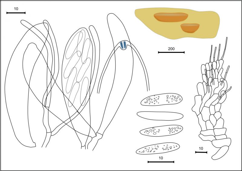
There are description and the pictures of the specimen from Er. russeolum (#4187).
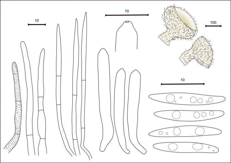
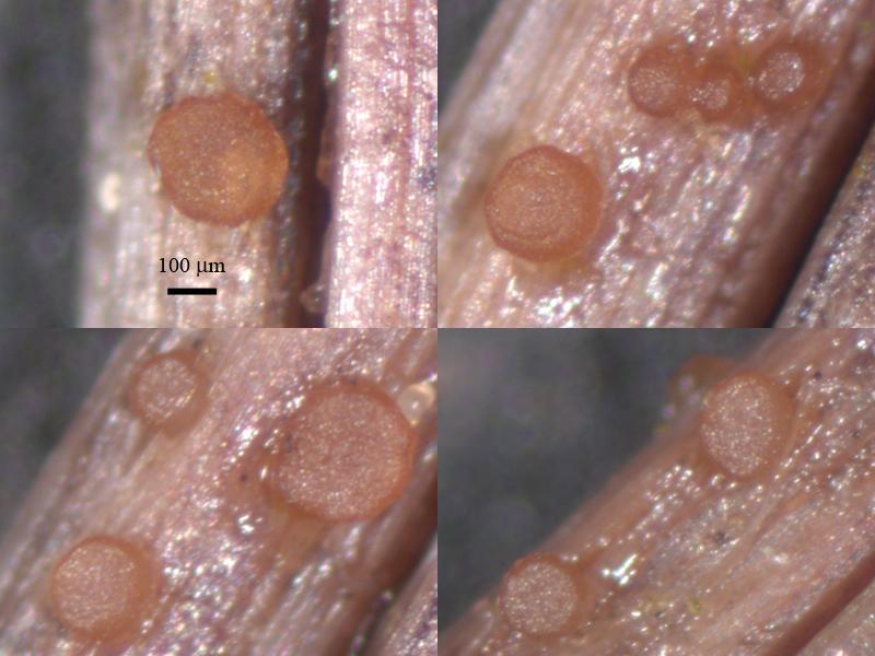
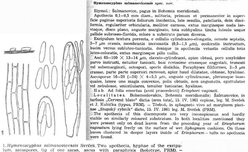
Apothecia discoid, sessile, thick, orangish, 220–320 mk in diameter, about 40 mk thick.
Excipulum from textura prismatica, cells with thickened walls, end cells cylindrical, obtuse; asci clavate, with obtuse-conical tip, without clamp, with small euamyloid pore, 75–94 x 11.4–15.6; paraphyses branched, about 1 broad in the middle, tips enlarged to 1.8–3.8, with some glueing incrustation at tips; spores ellisoid, with obtuse ends, 23 (18.9–26.3) x 5.9 (5.4–6.3) (n=18).?
I agree, specimen from Er.vaginatum looks similar except "hairs", probably they are some artefact, perhaps from drying since the substrate very thin and dries fast, i will check them once more to be sure.
Nina.
Di you look whether you had found this already in ealier years?
Zotto
thanks for you participation!
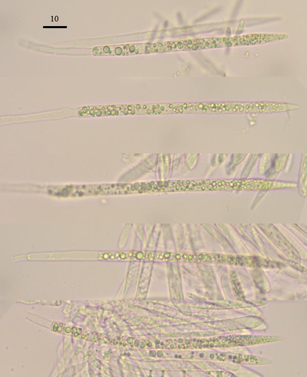
I had checked cottongrass litter yesterday again, and this Lachnum was met and the vital structures described (now i am enjoying the beauty of vital structures)),
It had no croziers.
No, unfortunatelly, not compared yet, just collecting and paying attention to better descriptions/photos.
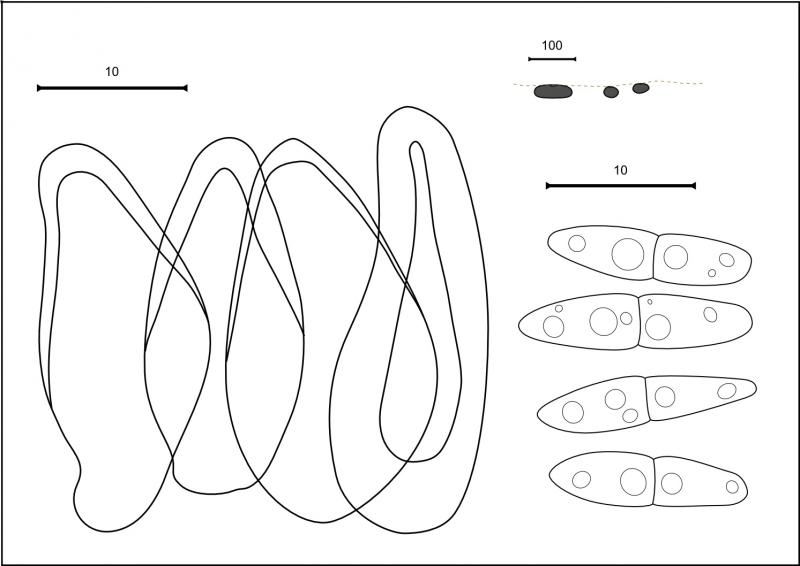
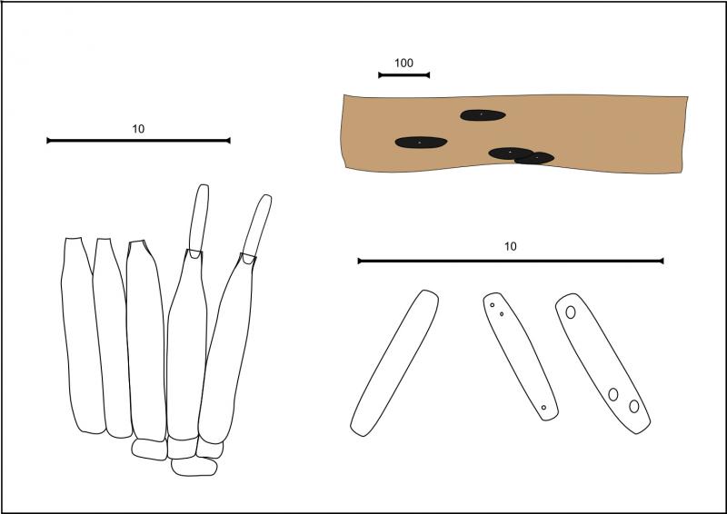
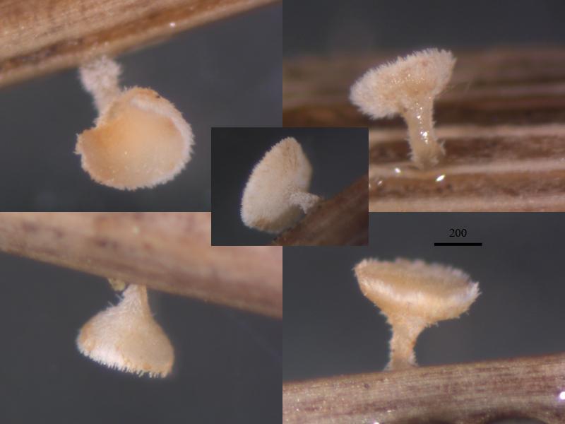
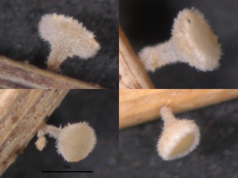
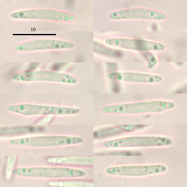
Lachnum (cf. juncinum) (#4268, not uploaded in Cubby)
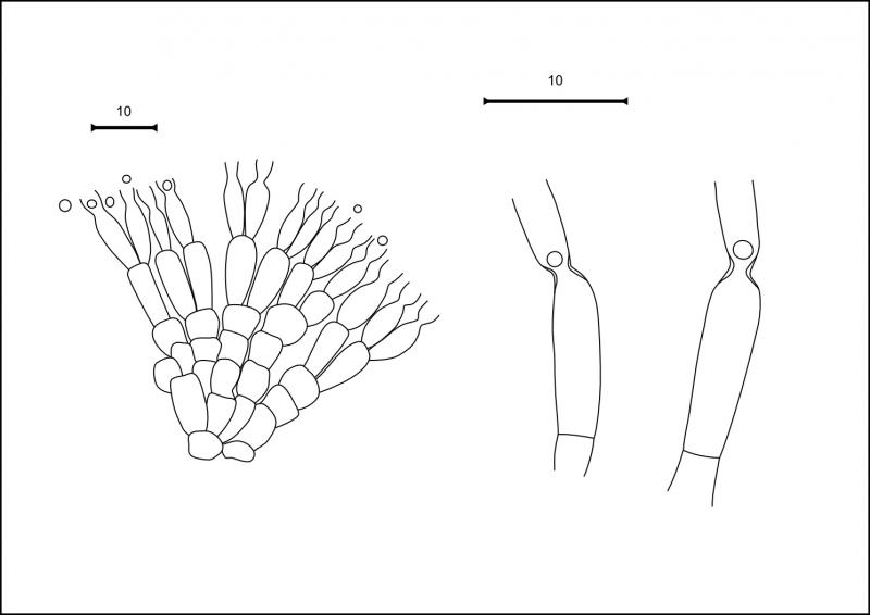
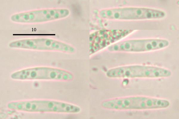
Apothecia cupulate, stipitate or short-stipitate, 430–750 km in diameter, 300–440 mk high; white and becoming yellowish or pinkish when dried.
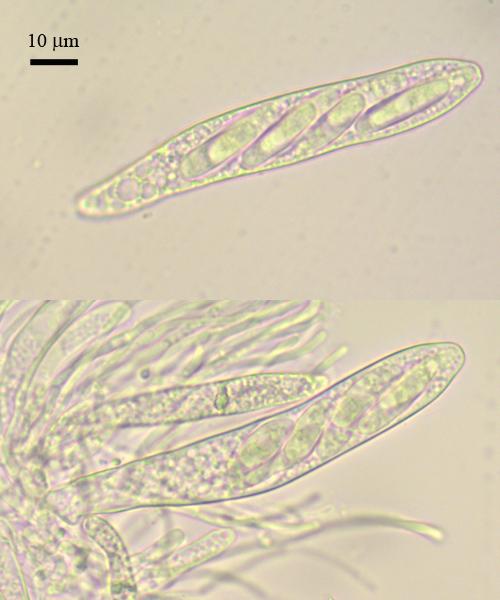
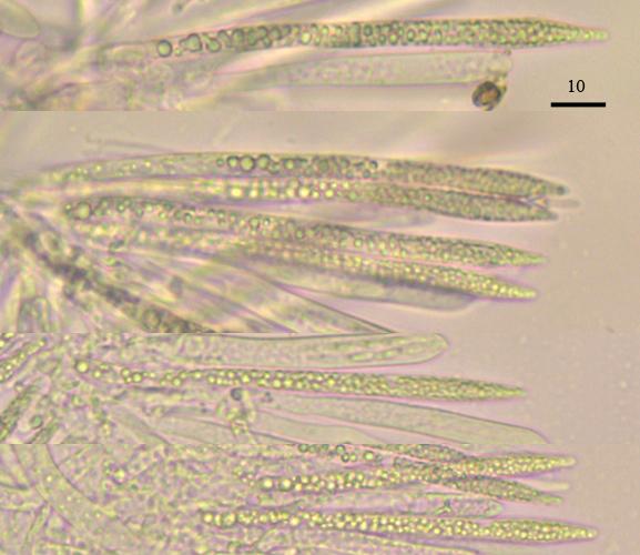
Hairs cylindrical, without enlargements, filled with abundant guttules, about 60 x 3.8; asci clavate, without crozier, about 67 x 7.6; paraphyses lanceolate, filled with abundant small vacuoles, about 82 x 4; spores fusoid, with several small oils, 15 (14–16.8) x 2.7 (2.5–2.8) (n=8).
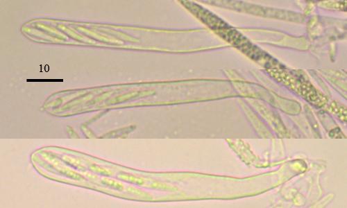
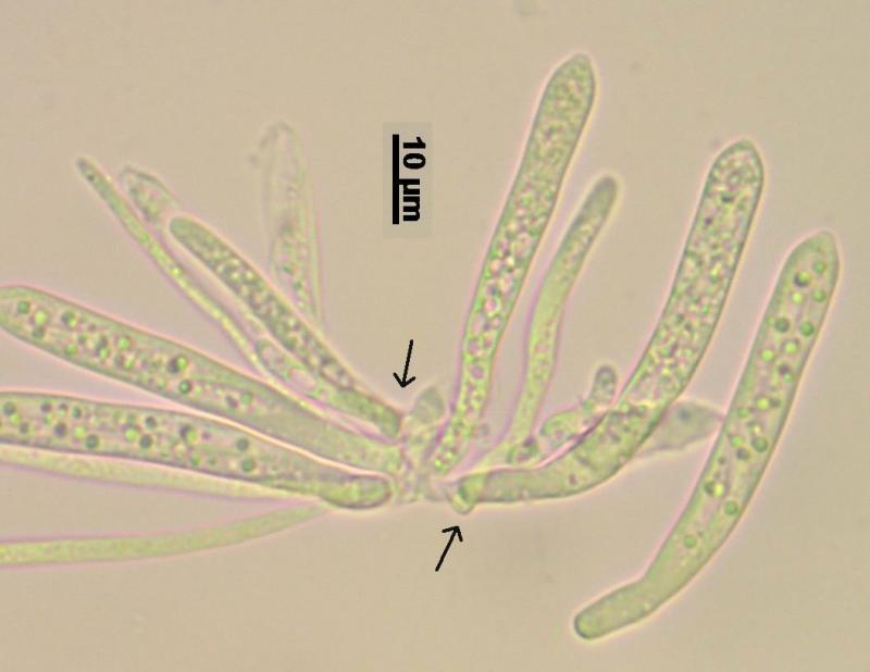
Now, are you sure with the absent croziers? Do you have aphoto where this is visible?
Here I think that your #4184 has croziers, but I did not seek thoroughly through your photos (Cubby is too slow, and I did not download all).
Zotto
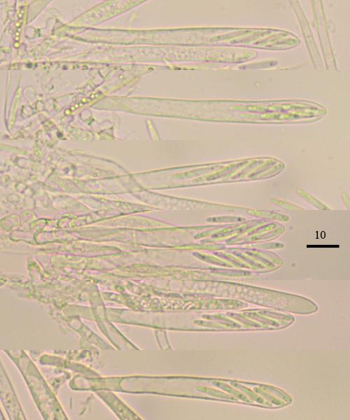
a had collected a larger collection of this species from several points, because i was thinking, probably different species may be there, with and without croziers.
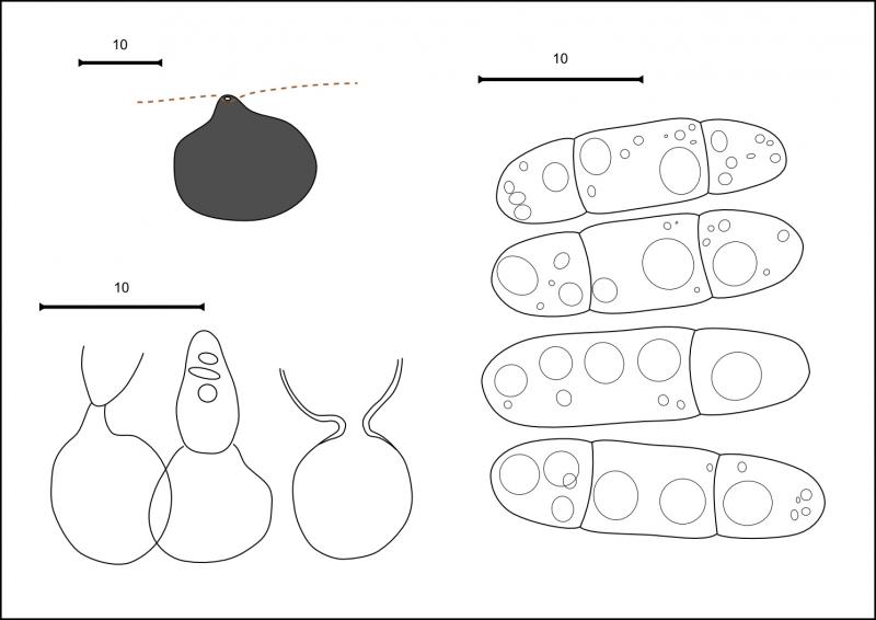
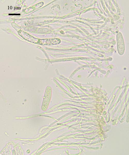
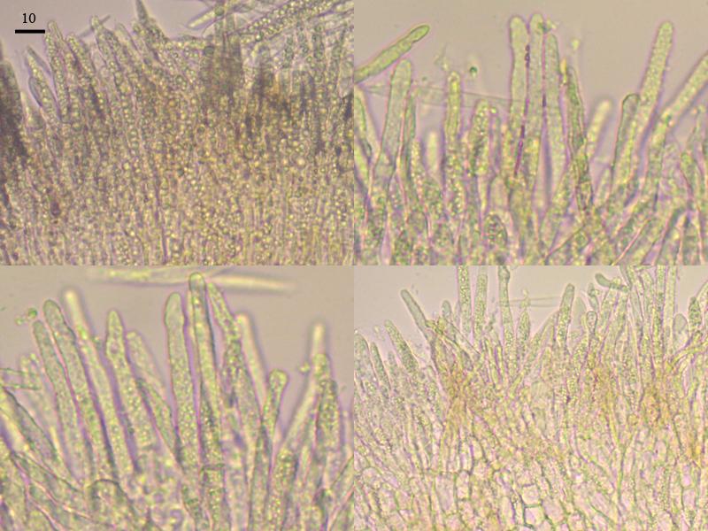
But i should correct my mistake, croziers are constantly seen in all collections, including re-checked previous one. Could be that the crozier become less seen in older (dead) asci? - probably that is why i am overlooking it.
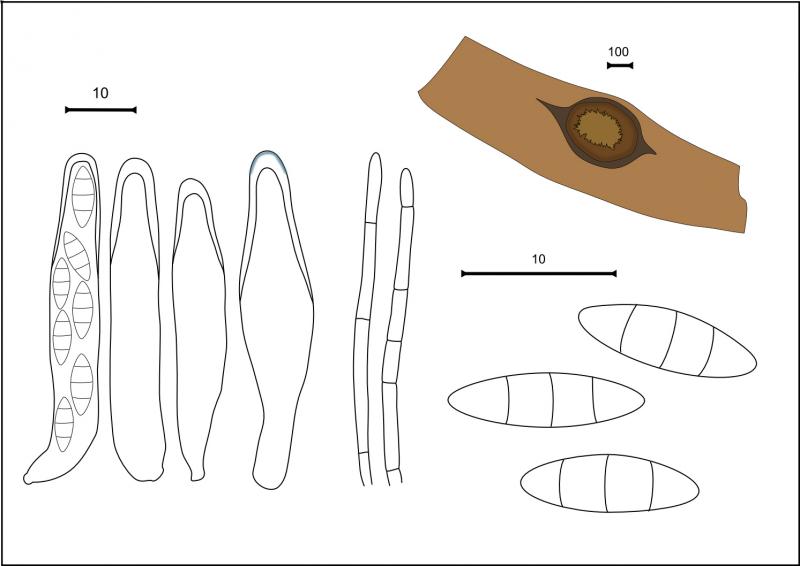
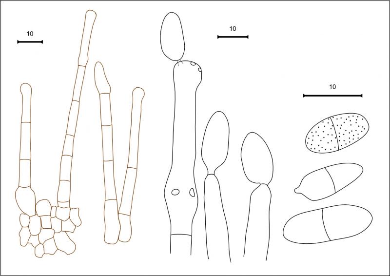
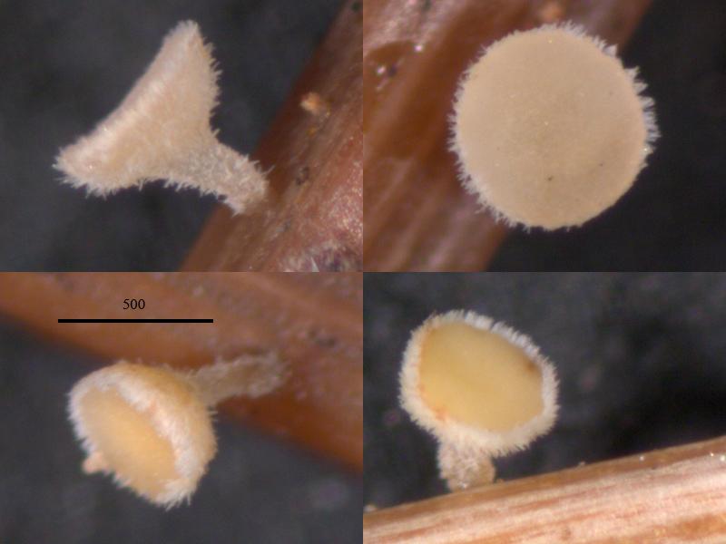
Spores could be narrower, there the description of the last collection (#4277):
Hairs cylindrical, without enlargements, segmented in lower part, with many small vacuoles, mean size 63 x 4, becoming brownish in exsiccates; paraphyses cylindrical, lanceolate in upper part, 2-3 segmented at base, mean size 117 x 2.8 (shape changes with age, old becoming many-septated, broader), filled with abundant VBs; spores fusoid, with several small oils, 14.3 (12.4–16.7) x 2.2 (1.9–2.5) (n=20).?
Nina.
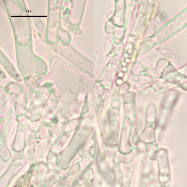
In overmature material croziers are difficult to see. The dead state may require mounting in KOH and staining with Congo, then the feature is usually very well visible. In fresh the feature is often seen at a glance.