
03-06-2026 19:45
 Miguel Ángel Ribes
Good afternoonI'm completely baffled by this suppo

03-06-2026 14:39
Thomas FlammerApothecia yellow, glassy-transparent, 80 - 120 ymS
16-03-2014 13:39
 Enrique Rubio
HI to all I'm looking for B. Hein's article on Wi
02-06-2026 14:33
 Nicolas VAN VOOREN
Hello.I'm searching for a PDF copy of the followin
18-10-2022 00:12
Valencia Lopez Francisco JavierHola amigos/asRecientemente encontré esta colecci
02-06-2026 17:58
Louis DENYBonjour forum, Sur feuille de Populus tremula, en

In a mixed wood with Quercus, Corylus, Alnus, Populus, Prunus, Ulmus on sandy soil. Found in several spots, near different herbs (among them Anemone).
I have not studied carefully the specimens in the first days, so part of the microphoto are taken after two weeks and are not very clear. The apothecia are dry now.
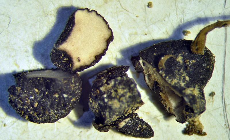
The sclerotia are black, rounded but always flattened. The sterile surface is delicately furfuraceous, this should be typical of S. sclerotiorum.
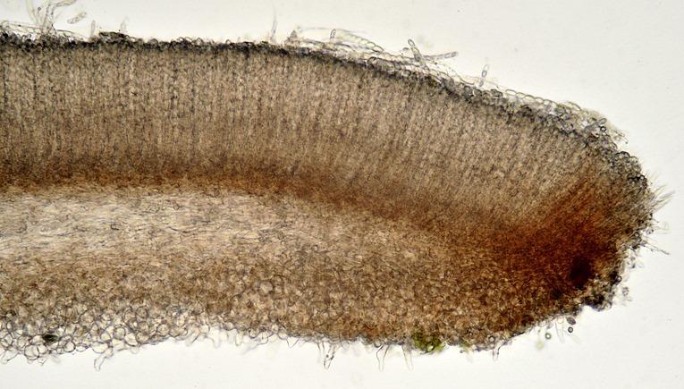
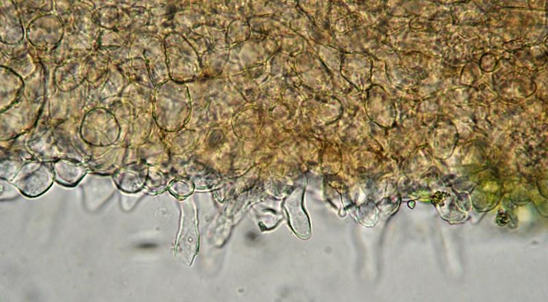
Ectal excipulum a textura globoso-angularis, non gelatinized (or not strongly so), apparently inamyloid but if the section is squashed in some areas appears some blue area (it seems some juice, not the walls to bluish) in iodine. The very outermost cells are elongated, thin-walled, somewhat prominent (that's why the surface is furfuraceous).
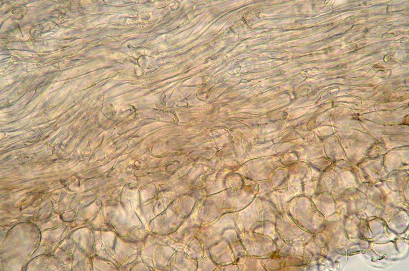
Medullary excipulum a textura intricata of rather thick, hyaline hyphae, mostly arranged radially.
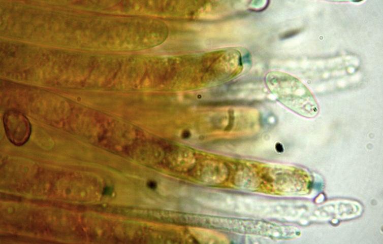
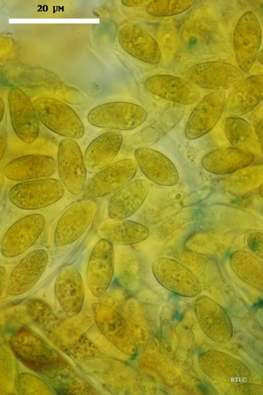
Asci with 8 spores all of the same size, 140-170 µm long, pore walls amyloid.
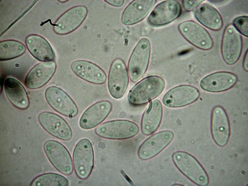
Ascospore roughly ellipsoid (not exactly symmetrical), with 2 to 4 very small (1 µm) droplets, 12-15x6-7,5 µm.
I think the spores are larger than normal for S. sclerotiorum, what do you think?
Thanks
Mario
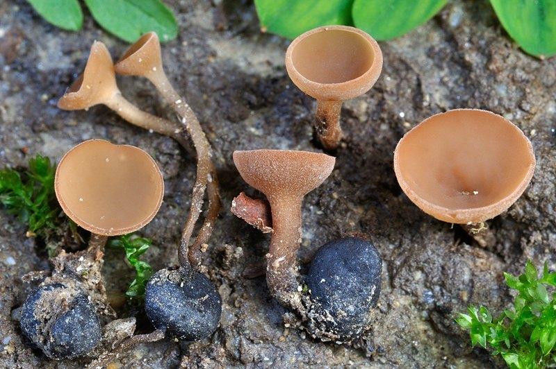

What is the diameter of your apothecia? There is a problem with Dumontinia tuberosa which has also which has also 2-nucleate spores, and a close relative with 2-nucleate spores. Both are about 10-20 mm diam., more cup-shaped and with often very long stalks, while S. sclerotiorum around 5-15 mm.
Zotto
Yes, I know I should have to observe the nuclei when it was fresh, but I didn't, sorry. Maybe they are still visible in dry specimens with Giemsa or acetic carmine...?
Maybe I can try to return on the spot to seek for some other apotecia, the collection was made three weeks ago...
I know D. tuberosa well enough to exclude it. These apothecia were rather small, not more than 10-12 mm; I found the first ones under a hazelnut so my first idea was an amenticolous Ciboria but then I have seen the sclerotia...
Usually D. tuberosa is bigger, darker, less regular (in the mature ones) and the sclerotia are more tuberoid, in my specimens I noted all sclerotia were round and flat.
Anemone was present in the wood but it was close only to some apothecia, the most were spread in the area near rather different herbs so the fungus should not be so host-specific.
Regards
Mario
Staining methods on dead material are rather complicated, I have no experience.
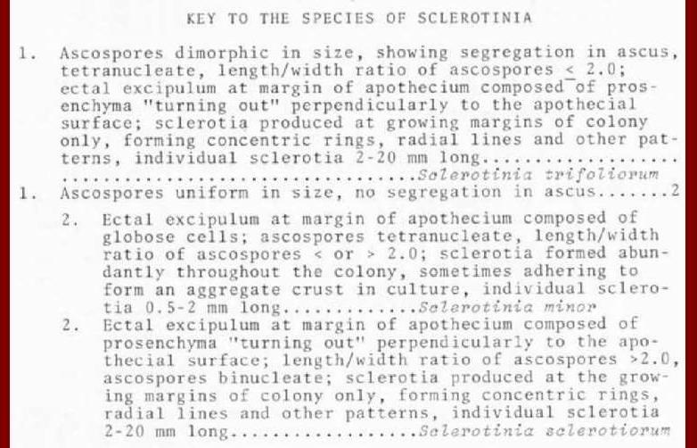
Then there is only the possibility of Scl. minor which is binucleate.
Zotto
actually Kohn 1979 says that
S. sclerotiorum is 2-nucleate and
S. minor 4-nucleate
Therefore it is not easy to separate between sclerotiorum and the large, Dumontinia-like species on Ficaria and Corydalis.
Zotto

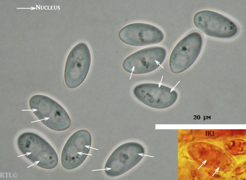
I suggest to try first with IKI before trying with any aggressive media. Maybe you can mix it with a bit of water if your IKI has too much iodine in order to see them better. If not the colour will be saturated. Spores may still be alive if little time has passed and you have done a normal drying. Anyway maybe the empty space taken up by the nuclei can still be seen as an empty area so you can estimate how many nuclei they were.
Sclerotinia sclerotiorum: 2 nuclei
Sclerotinia minor: 4 nuclei
I attach spores of S. sclerotiorum in water and IKI. I agree that spores are a bit wide to fit in S. sclerotiorum, moreover if you have rehydrated and after taken the measurements. But also take in consideration overmature spores which are not uncommon in this genus. Then abnormal spores can be seen, though not in your microphoto about spores. Can you provide a scale for that one? In my collections I never found over 7 µm wide.
My idea of what S minor is:
http://www.ascofrance.fr/search_forum/15005
Cheers,
Raúl
PD: I see Zotto has corrected in the meanwhile
Dumontinia tuberosa has 2 or 4 nuclei ?
I thought it was 4 nuclei. I have some collections with 4 nuclei on Anemone nemorosa.
Regards,
Daniel
I tried some hours ago to observe the spores of the collection with Giemsa, and in my amazement I have seen almost all the spores were germinating in the asci, so I think no possibility to understand exactly how many nuclei they contained. I can try again with other apothecia and with IKI, maybe tomorrow.
Zotto if the only choice is between S. minor and S. sclerotiorum, the ectal excipulum clearly indicates the latter... No need even to see the micro, the macrophoto (central specimen) should be enough...
But Kohn's key, is not too simplified?!?!
Regards
Mario
I can only speak about tuberosa/sclerotiorum. The excipular differences that Kohn reports and uses as diagnostic I could not really verify in several of the specimens that I had studied of both species.
Zotto
Maybe I have made a HUGE error. I have to work a little on that. 8-|
I was afraid to have considered as a Sclerotinia, something different.
Returned to the spot, while I was searching for the apothecia, I tried to find some relations with the roots of the host to identify it. The sclerotia are always superficial, the ascomata are easy to collect without to break the stipe, and I was not able to find any relations with the roots. Moreover, they are usually in the spaces between the herbs and never close to their stems. So I considered the possibility that the sclerotia arose from some kind of plant remnants in the litter more than from living roots; and I tried to identify all the leaves present in the litter. In addition to the trees I listed above, I found some leaves of Acer, some fruits of Castanea and, close to a cluster of 4 apothecia, a pod of Robinia. A Robinia pseudoacacia tree was just over my head, in fact.
I found two sclerotia on the surface of the ground, without apothecia.
And seeking for some other ones, I realized that there were many Robinia seeds, those little dark brown "beans"... rounded... and flattened(!) of the same size of the sclerotia...! Were they really sclerotia, or maybe stromatized seeds? That should change everything.
OK, at home however I cut some sclerotia, and they are exactly... sclerotia with a black rind and a white medulla, without any remnants of plants at the inside.
So I returned to the observation of the nuclei, this time in the living spores they were not difficult to see (in Lugol). The spores are binucleate so I think Sclerotinia sclerotiorum can
be confirmed. Maybe I will measure some others spores and try a statistics.
Thank you all!
Mario
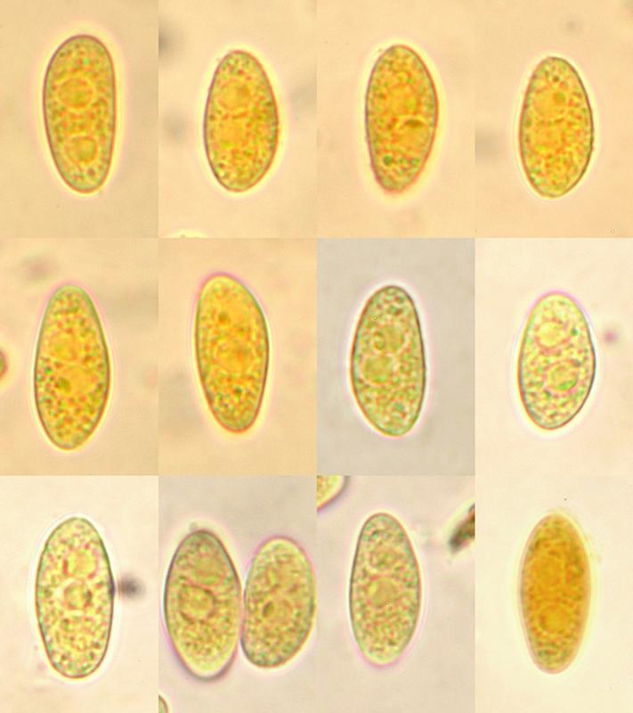
Just to compare, my Sclerotinia sclerotiorum spores range: 11,5-15 x 6-6,7
Cheers,
Raúl
Zotto