
26-04-2024 10:07
 Mathias Hass
Hello, Does anyone know what this is? Found on J
24-04-2024 21:54
 éric ROMERO
Bonjour, J'ai trouvé ce Lasiobolus sur laissées
23-04-2024 15:18
 Lothar Krieglsteiner
... but likely a basidiomycete. I hope it is o.k.
23-04-2024 13:17
 Edouard Evangelisti
Bonjour à tous, Je viens de récolter ce que je
23-04-2024 21:49
Ethan CrensonHello all, A friend recently found this orange as
22-04-2024 11:52
 Zuzana Sochorová (Egertová)
Hello,I made a loan of a collection of Microstoma
11-01-2022 16:36
 Jason Karakehian
Hi does anyone have a digital copy of Raitviir A (
22-04-2024 20:38
 Miguel Ángel Ribes
Good afternoon.Does anyone know this anamorph?It g

 Greetings AscoFrance!
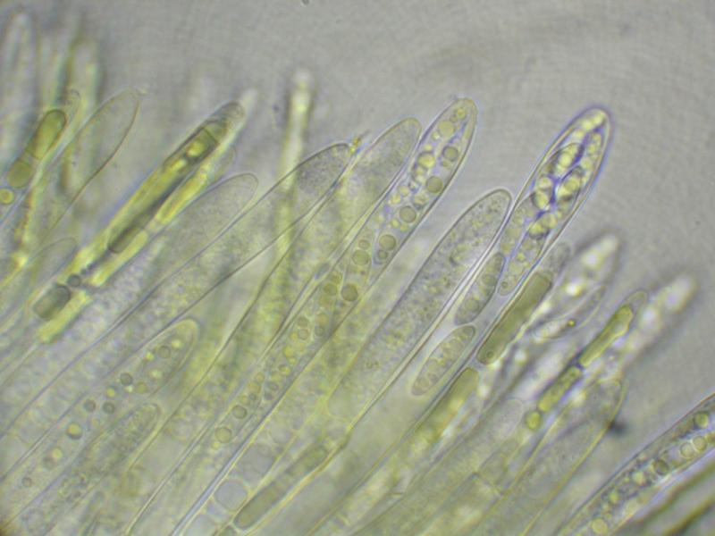
Here is a curious discomycete, field IDed to Ionomidotis by Paula DeSanto on the recent Peck Foray in Watkins Glen, New York. This particular find is from a mixed, predominantly hardwood forest within the Meads Creek State Forest. When I got a look at the dried material and field photos, I saw enough resemblance to my own Ionomidotis collection from North Carolina (http://mushroomobserver.org/174774) to consider the possibility, but upon preparing the material for microscopy we noticed that KOH extractable pigments (3% solution) were conspicuously absent. Can it still be Ionomidotis without this reaction? Perhaps this is a member of some other genus in the Encoelioideae?
Ascus tips inamyloid, despite appearing somewhat bluish in the micrographs. No paraphyses observed.
Spores:
9.5-14×=2.5-4.5?m (x=12.25×3.325?m, Q= 2.44-5.2?m, Qm=3.781?m, m=20, s=1)
13.5 x 3 ; 4.5
13 x 4 ; 3.25
13 x 2.5 ; 5.2
12.5 x 3.5 ; 3.57
14 x 4 ; 3.5
13 x 3 ; 4.33
9.5 x 3 ; 3.17
12.5 x 4 ; 3.13
13 x 3.5 ; 3.71
11 x 4.5 ; 2.44
13.5 x 3 ; 4.5
13.5 x 3 ; 4.5
10.5 x 4 ; 2.65
12 x 3 ; 4
12.5 x 2.5 ; 5
12.5 x 3 ; 4.17
9.5 x 3 ; 3.17
14 x 4 ; 3.5
11.5 x 3 ; 3.83
10.5 x 3 ; 3.5
Many thanks!
-Danny N.
PS: The images are all apparently too large for the site :( Please find them on Mushroom Observer here: http://mushroomobserver.org/218595

I am reminded of a Chlorencoelia, but the two species for which I have images, C. versiformis and C. torta) have distinctly amyloid asci. The spores would fit.
I am sure that the paraphyses would be seen when squashing the hymenium. If you had pictures from fresh material the genus Chlorencoelia would show a striking feature in the paraphyses (vacuolar bodies, see attach).
Zotto
Also, I believe the fact that the bottle of Melzer's used was labelled "Melzer's Replacement" may have something to do with the lack of observed blueing. Will use a more reliable reagent for the second set of micrographs.
These vacuolar bodies are a useful character at the family level. They are rather typical for the family Cenangiaceae as we now circumscribe it, but absent from the Cordieritidaceae which inbclude many ionomidotic species.
many thanks!