23-01-2026 21:50
Cameron DKI am looking for this please publication. is anyon

10-01-2026 20:00
Tom SchrierHi all,We found picnidia on Protoparmeliopsis mur

21-01-2026 16:32
Gernot FriebesHi,I need your help with some black dots on a lich

21-01-2026 16:48
Gernot FriebesHi,after my last unknown hyphomycete on this subst

20-01-2026 17:49
 Hardware Tony
I offer this collection as a possibility only as e

15-01-2026 15:55
 Lothar Krieglsteiner
this one is especially interesting for me because

17-01-2026 19:35
Arnold BüschlenHallo, ich suche zu Cosmospora aurantiicola Lite
• Reminiscent of Cudoniella, also not reddening, spores very long, exciple structure.
• It seems to be a good match for H. geogenus, the epithet was helpful too.
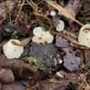
Habitat: Growing on the ground, on partially buried and decaying acorns and bud scales of Quercus, and possibly other woody debris, damp and muddy area under the tree, mixed (mostly) deciduous woodland, part of a large area of agricultural land being 'rewilded' (Knepp Estate), Low Weald, southern England, early November.
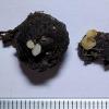
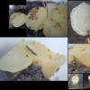
Apothecia: < ~3 x 6 mm, whitish to yellowish, rather dull and opaque appearance, yellowing with age, not reddening with trauma, gregarious to 5-caespitose, initially capitate-turbinate, then turbinate-pulvinate, eventually more appressed and pulvinate-discoid, superficial but emerging from debris, appearing sessile or with short and obconical stipe, receptacle and disc similar in appearance, disc initially concave and eventually plano-convex and often slightly umbilical.
Low magnification: Very short whitish hairs seen around the base of one, margin more noticeable when less mature, slightly felty, whitish and more reflective, often remaining slightly upturned but eventually reflexed, sometimes becoming slightly scalloped, sometimes eventually lobate, disc with slightly pruinose appearance.
Asci: Appear to be quite narrow, cylindrical-clavate, simple septa, rings bb, medium strength, Hymenoscyphus-type, deep dome-like thickening in flaccid immature asci, more subtruncate in more mature flaccid asci, mature asci abundantly discharging almost immediately in water (even straight from the fridge), thickening remaining everted, large collar-like appearance.
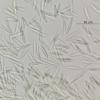
Spores: Ellipsoid-fusoid, approximately homopolar with the ends acute-rounded, although the apex is often more abrupt and acute, resp. the base is more elongated and rounded, asymmetric or slightly (sub)curved in profile view, flexible, multi-guttulate with OCI 5, many medium-size shadowy guttules (VBs?), transparent in IKI, some tiny dark LBs, apparently uninucleate, no setulae observed, one free spore with a central (transverse) septum.
Spores discharged from asci in water mount:
(28.7) 30.8-40.8 (41.9) × 5.5-6.1 (6.5) µm, Q = (4.8) 5.2 - 7.2 (7.5), N = 30, mean = 35.2 × 5.9 µm, Q mean = 6.0.
Paraphyses: Filiform (relatively narrow), Hymenoscyphus-type VBs at the apex, lowly refractive, faintly yellowish, osmetimes branching slightly irregularly and close to the apex.
Medullary: Text. intricata-porrecta, hyaline, deep, hyphae narrower and denser towards the subhymenium (some parallels with transition of ectal text.), more text. intricata, towards the base hyphae resp. broader and sparser, more text. porrecta.
Ectal: Somewhat disorganised, shallow, cells at the surface smaller and sometimes protruding, or irregularly clavate-lanceolate at the flanks, upper flanks more text. prismatica-angularis, parallel to the surface, surface of lower flanks and base text. angularis-globosa, below the surface lower flanks becoming more text. prismatica (perpendicular) in transition to the medullary, externally with pale yellowish pigment, stipe part not observed.
Marginal cells: Relatively short and narrow, cylindrical, apex rounded, 1-3? septa protruding, with some shadowy contents below the apex.


It is strange that IF does not have P. flavum as a synonym of P. monticola, although the entry was updated in 2022, is this synonymy published?
The morphological relationships between H. imberbis and Phaeohelotium (s.s./monticola group) are interesting. The macro and exciple structure between the groups is relatively similar but the paraphyses and VBs seem to be different. I can certainly see ecological and morphological similairities between H. geogenus and P. monticola.


 Hymenium-0026.jpeg