02-05-2026 12:42
Alain BRISSARDBonjour à tousJeuidi 30 avril dernier on m'a remi
02-05-2026 13:06
Pauline. PennaBonjour Please can someone help me with this id
01-05-2026 22:45
 Thierry Blondelle
Bonjour à tous, Une récolte sur bouse séchée d
28-04-2026 20:07
 Lothar Krieglsteiner
... on twig in the air at standing Ceratonia siliq
14-04-2026 05:32
Ethan CrensonHi all, A few weeks back a friend pointed out som
28-04-2026 20:33
Vitus SchäfftleinHello, I found Trochila ilicina on Ilex aquifoliu

30-04-2026 10:28
Rot BojanHello, by appearance I would say that I am dealing
27-04-2026 18:48
Tony MoverleyCollected 23rd April 2026, Norfolk, EnglandSwarms
27-04-2026 20:52
Lothar Krieglsteiner
Found on hanging tiwg of Olea europaea in dried-ou
 Hi everyone,
I got a fungus on bark from southern Italy, seemingly with no connection to the surrounding lichens. If it were lichenicolous, I would presume it to be a Buelliella. Has anybody any idea what this could be?
Ascomata sessile, constricted below, initially closed, later apothecioid, up to 0.5 mm diam., irregularly roundish, disc black but covered with a rusty pruina, margin prominent, densely rusty pruinose.
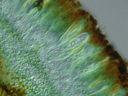
Hypothecium brownish, 55 µm high, hymenium hyaline below, brownish above, 120 µm high, epithecium brown, covered with dark brown granules, excipulum dark brown, up to 50 µm thick.
Paraphyses septate, sparsely ramified, 2–2.5 µm wide, hyaline below, brownish above, the upper cell sometimes enlarged up to 4 µm, brown in the upper half.
Asci clavate, 80–95 × 15–26 µm, apically thickened, with an internal beak, with a long stalk, 4–8-spored.
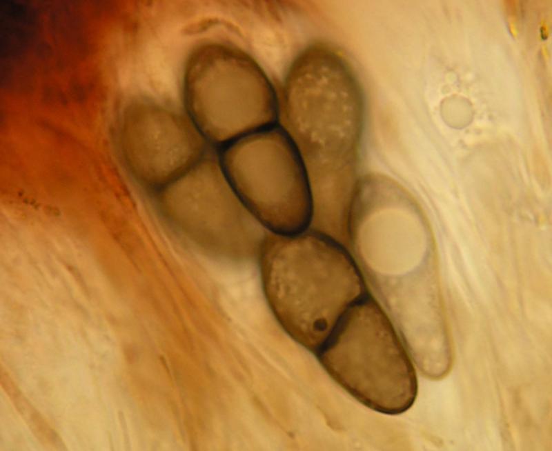
Ascospores 1-septate, grey, the upper cell rounded or slightly attenuated, the lower attenuated, narrower than the upper one, constricted at the septum, with one big guttule in each cell, surface ± smooth but appearing foveate, (20–)20.8–23.1(–24) × (8.5–)9–9.9(–10) µm, l/b = (2–)2.2–2.5(–2.6) (n = 20).
Pruina K+ violet, not dissolving. Hymenium above K+ grey, I+ reddish. Asci externally I/KI+ pale blue, no I/KI reacting apical structures.?