
17-03-2026 10:09
 François Freléchoux
Bonjour, Voici la description rapide d'un petit d
18-03-2026 13:09
 Khomenko Igor
I recently examined Celtis occidentalis branches
17-03-2026 19:41
 Bernard CLESSE
Bonsoir à toutes et tous,Pourriez-vous m'aider à
18-03-2026 17:22
Katarina PastircakovaHi there,I'm looking for the following literature:
19-03-2026 10:56
Thomas Læssøehttps://svampe.databasen.org/observations/10505643
27-02-2026 11:21
 Yannick Mourgues
Hi to all. Here is a specie that can may be relat
18-03-2026 18:42
Gonzalez Garcia MartaI have collected some lyre-shaped apothecia on the
27-11-2025 15:41
Thomas LæssøeSpores brownish, typically 4-celled; 26.8 x 2.4;
18-03-2026 11:52
Thomas Læssøehttps://svampe.databasen.org/observations/10493688
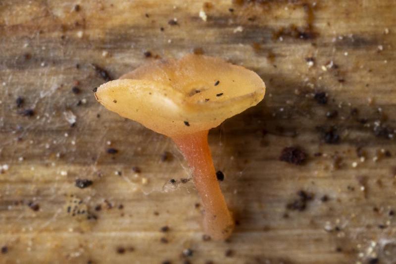
Cyathicula on Hemerocallis fulva
Johan Myhrer,
04-11-2018 18:24
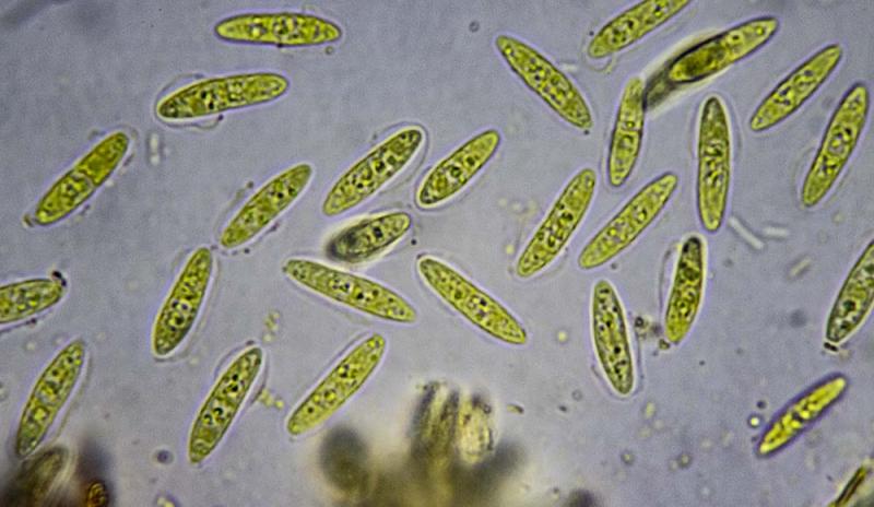
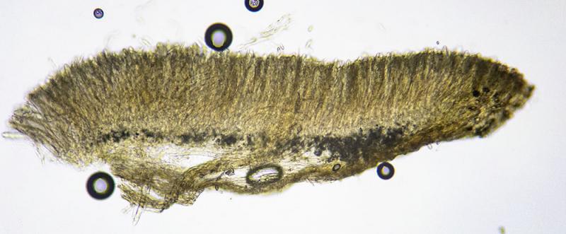
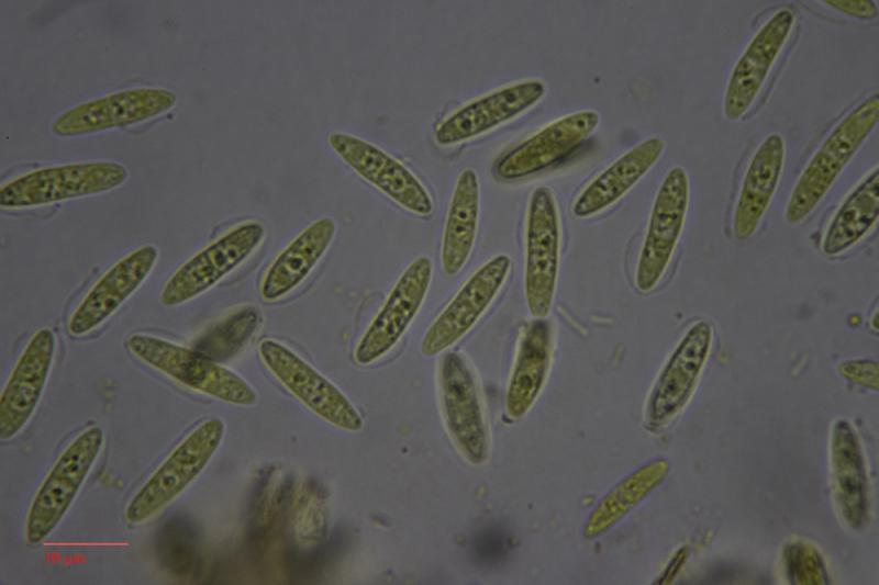
On dead flower stems of Hemerocallis fulva laying on soil in my garden (central Sweden 2018-11-04), I found these (along with typical Cyathicula coronata). Spores ~11x4µm, ascus apex IKI+ weakly blue. The latter difficult to observe because of "explosive" spore release when solution added. Could be overmatured C. coronata with septated spores? Measured spore size on coronata in this collection is ~21x4µm though. Thankful for guidance!
/Johan Myhrer

Hans-Otto Baral,
04-11-2018 18:34

Re : Cyathicula on Hemerocallis fulva
Dear Johan
I canot believe 11 x 4 µm. When I measure the spores are either 11 x 3 µm or 15 x 4.
The excipulum is strongly gelatinsed? Or could it be a Hymenoscyphus?
Explodung occurs in mature asci. usually there are immature ones and they are especially suitable for apical ring structure, of course in dead state when cell walls are thick.
As you observed abundant exploding, I assume that the figured spores are freshly ejected. This means that they were 1-septate within the living asci, a rare feature in Hymenoscyphus and also Cyathicula. But the guttulate paraphyses exclude a Calycina where 1-septate spores often occur.
Zotto
Johan Myhrer,
04-11-2018 19:14
Re : Cyathicula on Hemerocallis fulva
Might very well be Hymenoscyphus, any species that would fit? Few species recorded on Hemerocallis as it seems?
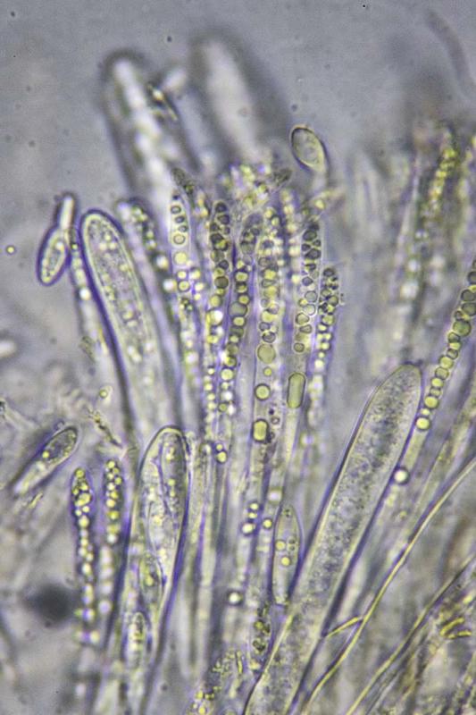
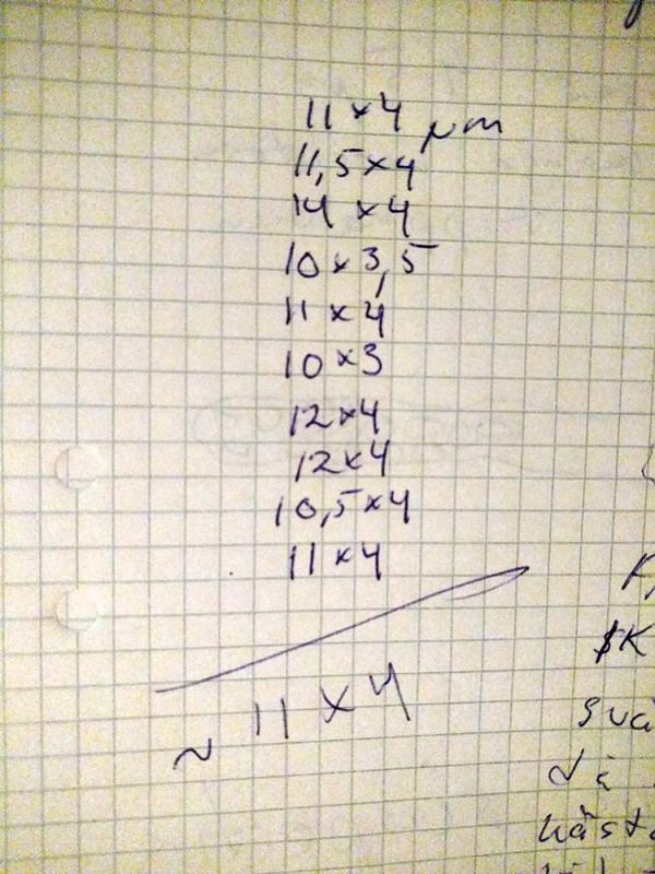
My notes from measurement of spores and picture of section:
Johan Myhrer,
04-11-2018 19:19
Re : Cyathicula on Hemerocallis fulva
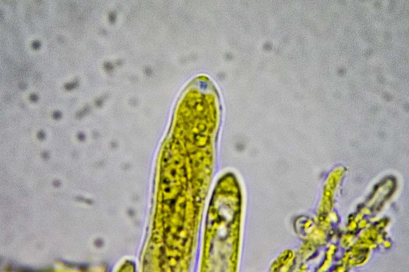
And ascus apex with IKI:
Johan Myhrer,
04-11-2018 19:33
Re : Cyathicula on Hemerocallis fulva
Second try with ascus:
Hans-Otto Baral,
04-11-2018 20:28
Re : Cyathicula on Hemerocallis fulva
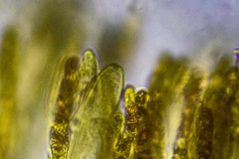
A closeup of the ectal excipulum would be necessary.
I do not suspect that the fungus is specific to a genus. Maybe to monocots. But I do not have an idea. The apical ring photos are splendid, they seem to exclude Hymenoscyphus because of the conical apex.
You don't have a scale to your spore photos?
I do not suspect that the fungus is specific to a genus. Maybe to monocots. But I do not have an idea. The apical ring photos are splendid, they seem to exclude Hymenoscyphus because of the conical apex.
You don't have a scale to your spore photos?
Johan Myhrer,
04-11-2018 20:50
Re : Cyathicula on Hemerocallis fulva
No scale to measurement on photos cause I photo through trinocular microscope and measure with scaled (calibrated) eyepeace :-/
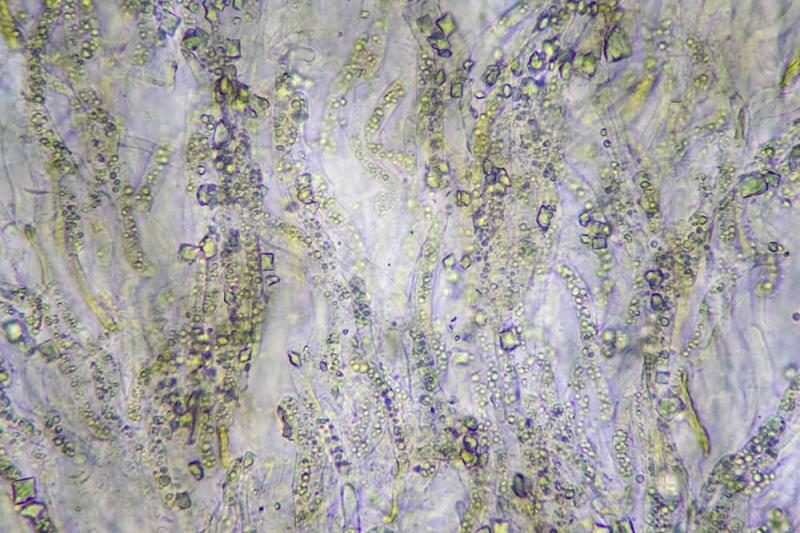
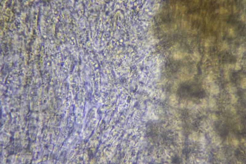
Ah, ectal excipulum:
Hans-Otto Baral,
04-11-2018 21:15
Re : Cyathicula on Hemerocallis fulva
You could easily photograph a calibration slide and prepare a scale to be included in a photo. That would enable control over measurements.
Your excipulum photo is great, it shows undulating guttulate living cortical hyphae and crystals - all typical of Cyathicula. The proper excipulum is below, however, and that could be gelatinized or not.
C. dolosella is a possibility, that has 1-septate spores in the living asci, *11-17 x 2.4-2.9. Ap margin with fine teeth
Zotto
Johan Myhrer,
04-11-2018 21:32
Re : Cyathicula on Hemerocallis fulva
Thank you for guidance Zotto! I will prepare a "calibration-picture" 'til next time. Have to give this Cyathicula a rest now though...
Johan Myhrer,
06-11-2018 15:11
Re : Cyathicula on Hemerocallis fulva
Tried a new method for spore measurement and it seems as I have to use this one in the future. Photoshop have a great; but a little complicated; measurement tool that I were not aware of!
Zotto; you where right about my measurements beeing to short, they are infact 12-16µm. So C. dolosella seems to be the one (Crocicreas dolosellum according to Swedish checklist).
/Johan Myhrer
 Cyathicula-spores-0001.txt)
Hans-Otto Baral,
06-11-2018 16:00
Re : Cyathicula on Hemerocallis fulva
O.k., and did you measure in Lugol or water? The nice photo in water must have another scale, can you find out that? because I fear that in Lugol the spores are already smaller (narrower).
I finally think that it is not dolosella. But the ectal excipulum is very difficult to squash?
I finally think that it is not dolosella. But the ectal excipulum is very difficult to squash?
Johan Myhrer,
06-11-2018 17:02
Re : Cyathicula on Hemerocallis fulva
Yes the excipulum is quiet tough. Measurement of spores in water as in attached files.
Hans-Otto Baral,
06-11-2018 17:42
Re : Cyathicula on Hemerocallis fulva
So it is mainly 14-15 x 3.8-4 µm.
Spore size of C. dolosella in Dennis 1978 is 12-15 x 2-2.5 (probably in dead state).
I think it is another species. The excipulum is not very clear, to me it looks thin-walled though difficult to separate. Cyathicula has 1-2 µm thick intercellular refractive gel.
Spore size of C. dolosella in Dennis 1978 is 12-15 x 2-2.5 (probably in dead state).
I think it is another species. The excipulum is not very clear, to me it looks thin-walled though difficult to separate. Cyathicula has 1-2 µm thick intercellular refractive gel.
Johan Myhrer,
06-11-2018 17:47
Re : Cyathicula on Hemerocallis fulva
I could not get the excipulum any clearer, this picture represents very well my observation through the eyepiece...