21-03-2026 15:13
Lepista ZacariasHello everyone, Does any one know of any literatu
20-10-2017 09:23
Garcia SusanaEste otro crecía en el mismo trocito de madera qu
20-03-2026 16:16
 Edvin Johannesen
These 0.5 mm diam. acervuli were breaking through
19-03-2026 19:34
 Filip Fuljer
Hello everyone,a few days ago I collected this str
19-03-2026 18:25
 William Slosse
Good evening everyone, On 18/03/26 I found a few
17-03-2026 10:09
 François Freléchoux
Bonjour, Voici la description rapide d'un petit d
19-03-2026 17:50
 Enrique Rubio
Hi to everybodyThese thiny, blackish pseudothecia
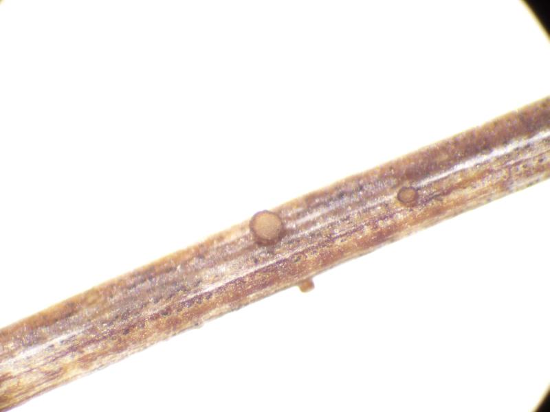
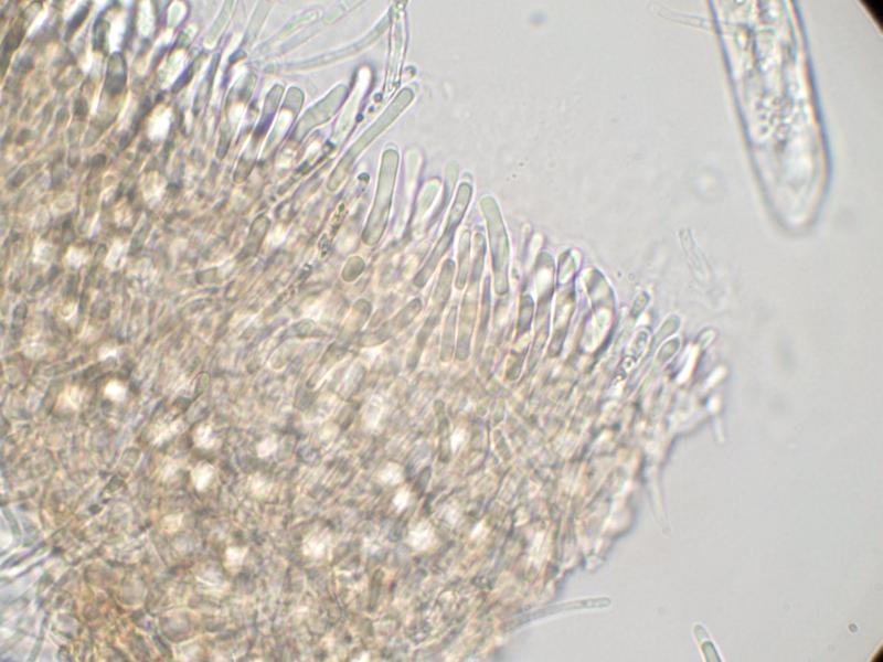
Apothecia sessile; cup-shaped; ca 300 µm diam.; hymenium pale grey; exterior brownish orange.
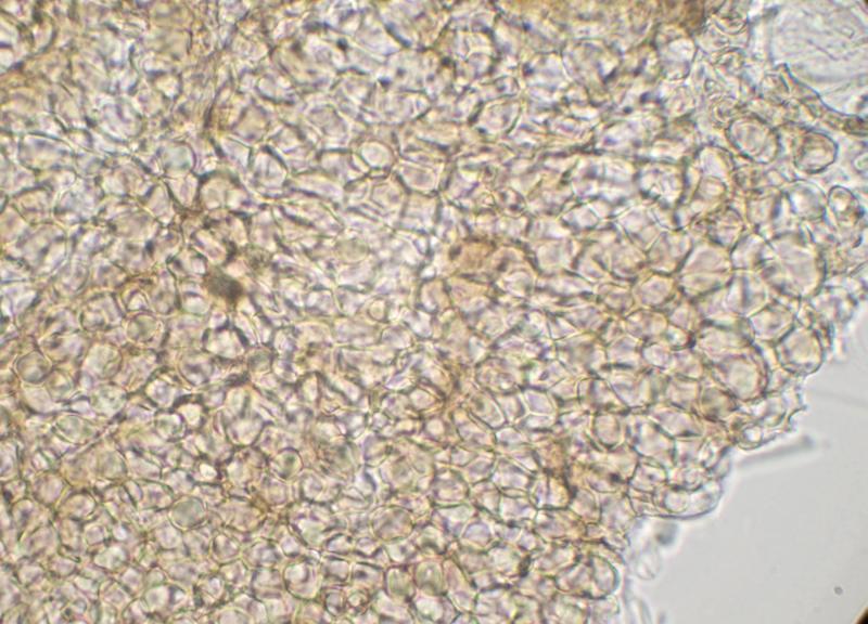
Excipulum brown textura globulosa.
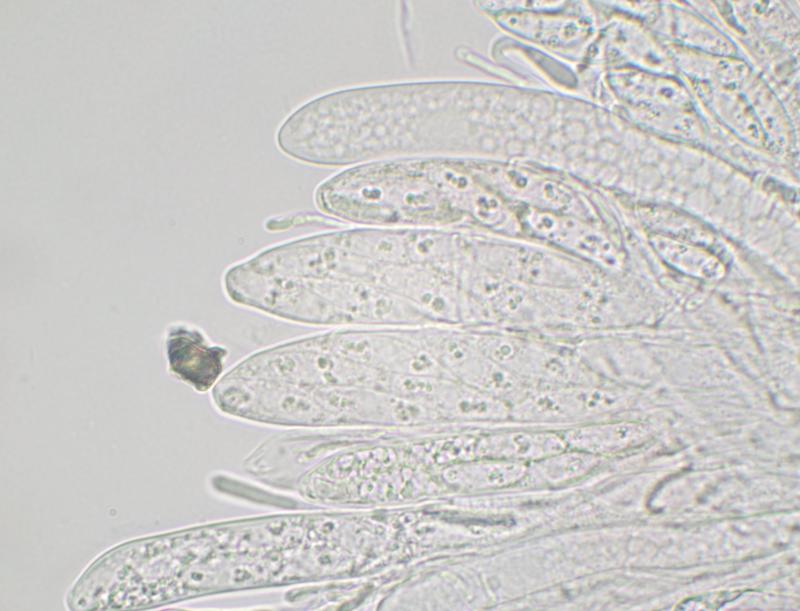
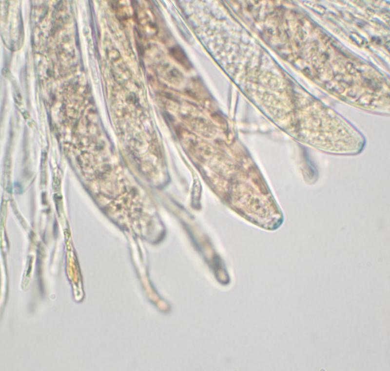
Paraphyses narrowly cylindrical (2-2.5 µm wide), sometimes swollen at apex; sometimes branched; cylindrical refractive VB in upper part.
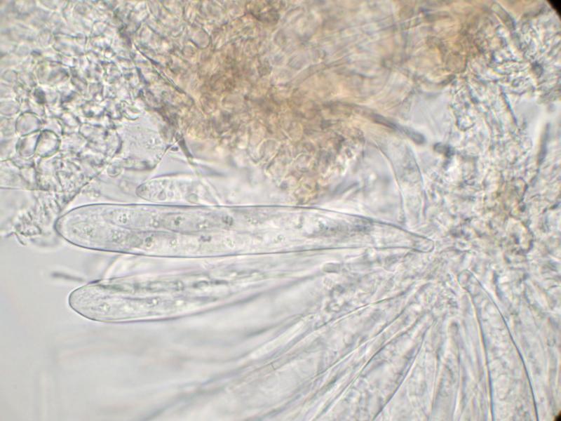
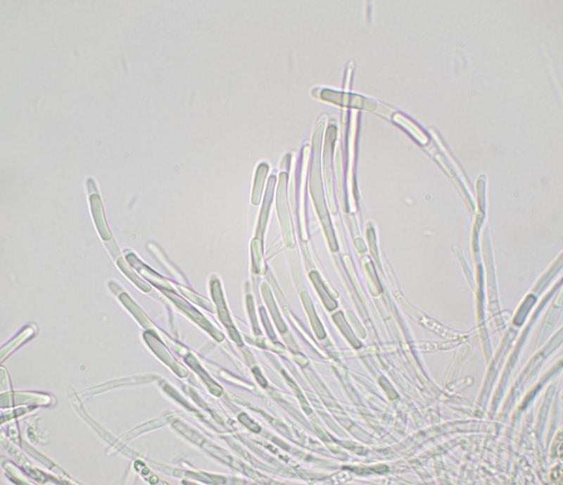
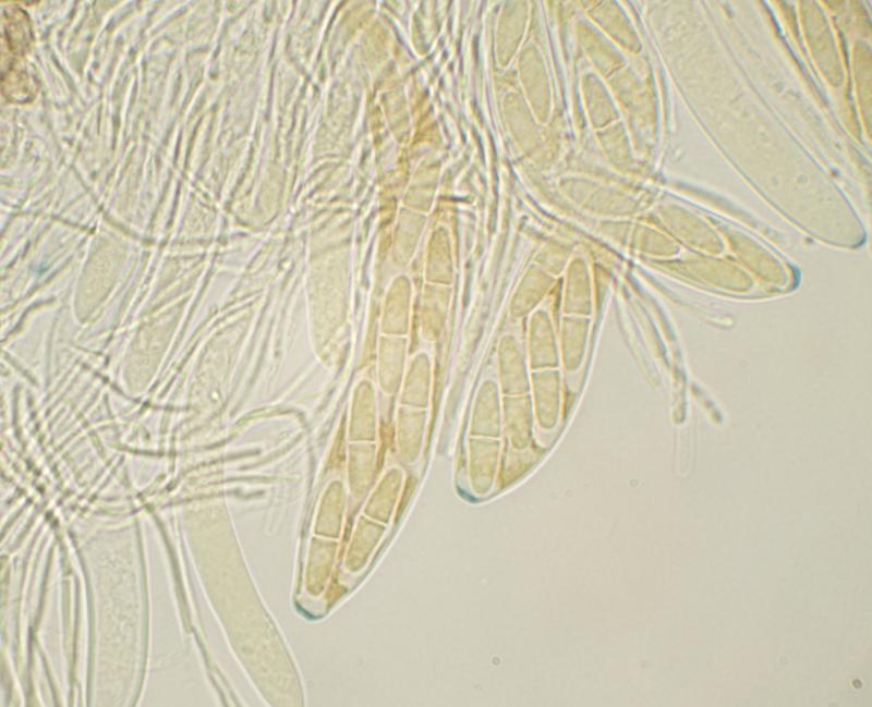
Asci clavate; ca 100-110 x 13-16 µm; 8-spored (biseriate); IKI+ blue; with shallow apical ring.
Ascospores fusiform; hyaline; free spores 21-23 x 6 µm; mostly 1-septate, but free spores sometimes 2-septate; scattered small OBs, mainly near ends of spore.
This seems to resemble Nimbomollisia (Niptera) eriophori. I didn't notice gelatinous sheaths on the spores when examining the specimen but the image of spores in the ascus in MLZ seems to show some sort of gelatinous structure at the ends of the spores. Some of the paraphyses have swollen apices but this feature isn't as well developed as I would have expected in Nimbomollisia.
I'd be grateful for a second opinion.
Thanks
Marcus
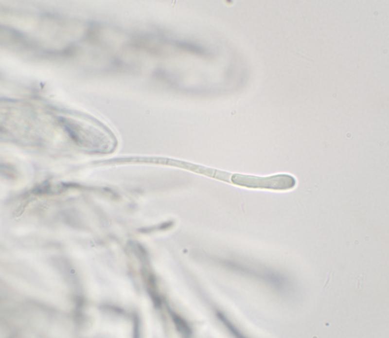
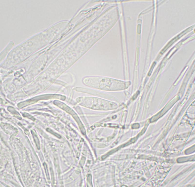


There were very few free spores. Spores in the asci were difficult to see clearly (see images) but I couldn't see any obvious caps or sheaths.